Das Behandlungsmodell Hospital at Home in der Schweiz

Hospital at Home als international anerkanntes ­Versorgungskonzept

Unter dem Begriff Hospital at Home (HaH) versteht man eine Versorgungsform, bei der somatisch akut erkrankten, hospitalisationspflichtigen Patient/-innen eine spitaläquivalente Behandlung zu Hause statt im Spital ermöglicht wird. HaH ist nicht für alle spitalbedürftigen Patienten geeignet, aber für eine ausgewählte Zielgruppe, bei der eine sichere, medizinisch-pflegerische Versorgung und Behandlung im häuslichen Umfeld möglich ist.

Mit dem HaH-Ansatz soll die Gesundheitsversorgung wieder näher zu den Menschen gebracht werden und sowohl der Genesungsfortschritt (u. a. besserer Schlaf durch gewohnte Umgebung und Versorgung durch Angehörige) als auch die Sicherheit von Patient/-innen (u. a. Reduktion des Infektionsrisikos, Vermeidung von Delir, geringerer Schlafmittel- und Sedativaverbrauch, weniger Stürze und Frakturen) erhöht und der Ressourcenverbrauch im Gesundheitswesen reduziert werden (1, 2). Internationale Studien zeigen zudem, dass die Autonomie der Patient/-innen zu Hause gestärkt wird und dadurch deren Unabhängigkeit leichter aufrechterhalten oder wiederhergestellt werden kann (3, 4), was nachhaltig zur Gesundheitsförderung beiträgt.

Darüber hinaus fördern die Patientenzentrierung und die intra- und interprofessionelle Zusammenarbeit in der neuen Versorgungsform eine Steigerung der Berufszufriedenheit oder -attraktivität (5, 6), wodurch man sich eine längere Berufsverweildauer des medizinischen und pflegerischen Personals erhofft.

International, insbesondere in den USA, Israel, Spanien und UK, ist HaH ein fixer Bestandteil des Gesundheitswesens (7). Eine Cochrane-Review, basierend auf 20 randomisierten Studien zu HaH, wies eine hohe Sicherheit der Patient/-innen, niedrigere Komplikationsraten als im klassisch stationären Setting, geringere Behandlungskosten und eine hohe Zufriedenheit der Patient/-innen aus (8). In der Schweiz stellt HaH ein neues Versorgungsmodell im Gesundheitswesen dar, welches in einer zunehmenden Anzahl von Projekten angeboten und erprobt wird (9).

Die Klinik Arlesheim gehört zu den Pionieren von HaH in der Schweiz, wo die neue Versorgungsform seit Januar 2023 in einem Team von Ärzt/-innen und Pflegefachpersonen implementiert wurde (10). Nach einer Machbarkeitsprüfung und Entwicklungsphase in den Jahren 2020 bis 2022 wurden von Januar bis Juni 2023 die ersten Patient/-innen im Rahmen von HaH behandelt. Seit Juni 2023 wird ein relatives feststehendes Konzept im Rahmen einer Pilot- und Evaluationsphase angeboten, welches durch den Kanton Basel-Landschaft unterstützt wird. Seit Beginn im Juni 2023 bis Ende Februar 2025 wurden ca. 350 Patient/-innen spitaläquivalent zu Hause behandelt.

Nachfolgend soll am Beispiel der Klinik Arlesheim ein Einblick gegeben werden, wie ein solches HaH-Programm praktisch funktioniert und welche Wirkungen und Möglichkeiten die neue Versorgungsform mit sich bringt. Weiterhin werden erste Ergebnisse hinsichtlich der Versorgungssicherheit, Behandlungsqualität und Zufriedenheit dargestellt.

Das Pilotprojekt an der Klinik Arlesheim

Motivation und Zielsetzung

Für das Team von Hospital at Home sind die medizinische Versorgungs- und Behandlungsqualität und die Sicherheit der Patient/-innen höchste Priorität. Das Angebot einer HaH-Behandlung ist freiwillig, und die Patient/-innen können selbst entscheiden, ob sie das Angebot annehmen oder wie sie herkömmlich im klassischen stationären Setting behandelt werden möchten. An- und Zugehörige werden im Rahmen der Behandlung miteinbezogen, beraten und angeleitet.

Parallel zur Versorgung der Patient/-innen entwickelt das Team von Hospital at Home gemeinsam mit dem Verein hospitales ein mögliches, helvetisiertes Konzept «Hospital at Home» weiter und widmet sich der Team- und Organisationsentwicklung mit modernen Arbeits- und Organisationsformen. Dabei setzt das HaH-Team in Arlesheim auf einen hohen Grad an Selbstwirksamkeit eines jeden Mitarbeitenden und arbeitet mit flachen Hierarchien und einer besonderen Meetingkultur, die eine Mitbestimmung und -gestaltung des gesamten Teams ermöglichen.

Praktisches Vorgehen

Team Klinik Arlesheim
Das HaH-Team besteht aus festangestellten, erfahrenen internistischen Fachärzt/-innen und Assistenzärzt/-innen und aus diplomierten Pflegefachkräften. Alle Teammitglieder sind mit der Versorgung internistischer Patient/-innen im stationären Setting vertraut. Eine enge Zusammenarbeit mit Therapeut/-innen, Spitex (spitalexterne Hilfe und Pflege), Hausärzt/-innen, Radiologieinstituten und Fachärzt/-innen (Konsile) besteht, und diese können situativ zugezogen werden.

Zuweisung und Patientenaufnahme
Die Zuweisung zur HaH-Behandlung der Klinik Arlesheim erfolgt in ca. der Hälfte der Patient/-innen direkt durch den Hausarzt und zu einem Viertel durch den Notfall. Eine geringe Anzahl der Patient/-innen wird aus stationärer Behandlung oder von anderen Fachärzt/-innen zugewiesen.

Eine Zuweisung in die HaH-Behandlung erfordert eine ärztliche Feststellung der stationären Behandlungspflichtigkeit. Anschliessend erfolgt durch das interprofessionelle Behandlungsteam eine pflegerische und ärztliche Einschätzung, ob eine HaH-Behandlung sicher durchführbar, technisch möglich, sozial erwünscht und medizinisch sinnvoll zu Hause machbar ist (Abb. 1). Die Evaluation dieser Kriterien, welche als Sicherheitsprüfung dienen, erfolgt kontinuierlich, beginnend bei der ärztlichen Zuweisung und endet mit einer abschliessenden Prüfung während der Aufnahmevisite im Zuhause der Patient/-innen. Erst im Anschluss wird ein erstes Therapiekonzept festgelegt. Weiter wird bei Bedarf das kontinuierliche Home Monitoring installiert und der/die Patient/-in wird in dessen Handhabung instruiert. Eine Aufnahme ins HaH ist von Montag bis Sonntag während 24 h möglich.

Eine Chance der neuen Versorgungsform bietet die Behandlung von besonders vulnerablen Patient/-innen in heilpädagogischen Einrichtungen sowie Alters- und Pflegeheimen. Es besteht die Möglichkeit, dass die Bewohner/-innen in ihrer Wohneinrichtung bei akuten Erkrankungen, die hausärztlich nicht mehr verantwortet und versorgt werden können, anstelle einer Spitaleinweisung durch das HaH-Team in der Wohneinrichtung behandelt werden.

Das Team von Hospital at Home kann je nach medizinisch-pflegerischen Behandlungsaufwand in der untersuchten Phase 4 bis 8 Patient/-innen gleichzeitig behandeln.

Behandlung
Das HaH-Konzept beinhaltet tägliche Visiten durch pflegerisches und ärztliches Personal. Dauer und Häufigkeit der Visiten werden in Abhängigkeit des klinischen Zustands sowie des Bedürfnisses des/der Patient/-in skaliert. Nach bisherigen Erfahrungen benötigen nur etwas über 30 % der Patient/-innen mehr als 2 Visiten pro Tag. Die Hälfte der Patient/-innen kann nach maximal 1 Woche entlassen werden.
Die Therapieintensität der Behandlung wird analog zu einem Spitalaufenthalt nach Bedarf des/der Patient/-in im Behandlungsverlauf angepasst und ständig reevaluiert. Das Equipment für Diagnostik, Therapie und Versorgung inkl. Medikamente, EKG (Elektrokardiogramm), Ultraschall, POCT(Point of Care Testing)-Labor sowie Notfallequipment werden durch das HaH-Team mitgebracht. Intensivere Untersuchungen, z. B. Magnetresonanztomografie (MRT), Computertomografie (CT), endoskopische Untersuchungen oder Echokardiografie werden, sofern notwendig, mit regionalen Gesundheitsdienstleistern durchgeführt.

Am Tag vor der Entlassung erhalten die Patient/-innen notwendige Medikamentenverschreibungen und einen Austrittsbericht. Zudem werden Termine zur weiteren ambulanten Anschlussversorgung, wie z. B. Physiotherapie oder den nächsten Termin bei dem/der Hausärzt/-in, vereinbart. Um einen nahtlosen Übergang in der ambulanten Weiterbehandlung zu gewährleisten, erfolgt die direkte Rücksprache mit dem weiterbehandelnden Team (Hausärzt/-innen, Spitex, sofern etabliert), welche auch den Austrittsbericht von der HaH-Behandlung bekommt.

Im Falle eines Notfalls oder eines dringlichen medizinischen Vorkommnisses haben Patient/-innen rund um die Uhr die Möglichkeit, das HaH-Team zu kontaktieren. Es besteht eine 24/7 h ärztliche und pflegerische Rufbereitschaft. Zur Gewährleistung einer maximalen Sicherheit der Patient/-innen erfolgt bei Bedarf ein 24/7 h telemedizinisches Monitoring der Vitalparameter (10). Nach den bisherigen Erfahrungen benötigen nur 10 % der Patient/-innen ein telemedizinisches Monitoring.

Komplikationen (z. B. Delir oder Stürze) sowie Spitalverlegungen während der HaH-Behandlung werden systematisch im Rahmen der pflegerischen und ärztlichen Betreuung erfasst.

Erste Ergebnisse

Nachfolgend werden deskriptiv die Ergebnisse aus einer retrospektiven Analyse aller 89 Patient/-innen, die zwischen dem 1. Juni 2023 und dem 31. Dezember 2023 im HaH der Klinik Arlesheim behandelt wurden, dargestellt.

Hospital at Home erreicht ein vielfältiges, ­geriatrisch geprägtes Patient/-innen-Kollektiv

Das Alter (Standardabweichung, SD) des HaH-Kollektivs lag durchschnittlich bei 73 (± 18) Jahren (55 % weibliche Patienten). Die im HaH behandelten Diagnosen verdeutlichen das breite Spektrum an Erkrankungen, welches in diesem Versorgungsmodell behandelt werden kann (Abb. 2).

Der Mittelwert des Charlson-Komorbiditätsindex (SD), welcher das generelle Mortalitätsrisiko basierend auf der Anzahl und Schwere der Begleiterkrankungen darstellt, lag bei 2.8 (± 2.2, Range von 0–10), was einem 1-Jahres-Mortalitätsrisiko von ca. 10 % entspricht (11). 53 % der im HaH behandelten Patient/-innen wiesen einen Selbstpflegeindex (SPI) unter 32 auf (bei einem mittleren SPI [SD] von 31.9 [8.1]) (12). Der SPI (Range von 0–40) dient als Marker für die Selbständigkeit der Patient/-innen, wobei tiefere Werte einen höheren pflegerischen Aufwand bedeuten und bei Werten unter 32 weitere Schritte zur Abklärung der poststationären Versorgung einzuleiten sind. Eine Studie in 1372 Patient/-innen, die notfallmässig in die Innere Medizin des Kantonsspitals Aarau aufgenommen wurden, wies einen mittleren SPI (SD) von 35.6 (6.0) auf (13).

Diese Werte demonstrieren, dass es sich bei dem behandelten HaH-Kollektiv um polymorbide Patient/-innen mit erhöhtem Pflegeaufwand handelte.

Hospital at Home ermöglicht den Verbleib in der ursprünglichen Versorgungssituation

Tab. 1 stellt die Veränderung der Versorgungssituation des Patient/-innen-Kollektivs vor und nach der HaH-Behandlung dar, wobei die Situation bei Austritt sich auf die endgültige Versorgungssituation der Patient/-innen bezieht, die bei Spitalüberweisungen erst später eintrat. Aus der Kreuztabelle wird ersichtlich, dass 78 der 89 Patient/-innen in ihrer Versorgungssituation verblieben. 6 von 55 (11 %) Patient/-innen, die sich vorher noch selbst versorgt haben, erhielten nach der Entlassung Unterstützung durch die Spitex. Nur bei 3 Patient/-innen kam es in Bezug auf die Betreuungsintensität der Spitex zu einem höheren Betreuungsbedarf als vor der HaH-Behandlung. 12 Patient/-innen wurden in einem Pflegeheim betreut und verblieben auch dort.

4 Patient/-innen verstarben, bevor sie die durch die HaH begonnene Versorgung beendet hatten, und 1 Patient starb nach vorheriger Verlegung in ein anderes Spital. Alle diese Patient/-innen befanden sich in einer palliativen Lebenssituation und wurden aufgrund dieser oder aufgrund einer zusätzlichen akuten Erkrankung zugewiesen. Zu einem unerwarteten Todesfall ist es nicht gekommen.

10 Patient/-innen wurden während der HaH-Behandlung in ein Spital überwiesen, was in 7 Patient/-innen aus medizinischen oder technischen Gründen (z. B. aufgrund einer Zustandsverschlechterung oder zur weiteren Dia­gnostik) notwendig war und in 3 Patient/-innen aus sozialen Gründen (häusliche Unterversorgung oder Wunsch der Patient/-innen) erfolgte. Zu einer Verlegung pflegebedürftiger Patient/-innen aus dem häuslichen Setting in die Kurz- oder Langzeitpflege kam es im HaH-Kollektiv der Klinik Arlesheim nicht. Ebenso wurde bei keinem/ keiner Patient/-in ein Bedarf für eine geriatrische Rehabilitation festgestellt.

Hospital at Home zeigt ein geringes Auftreten unerwünschter Ereignisse

Bei 3 Patient/-innen kam es unter dem HaH-Aufenthalt zu einem Sturz, was eine beobachtete Sturzrate von 4.1 Stürzen pro 1000 Pflegetagen ergibt. Laut verschiedener internationaler Studien, welche über die Sturzhäufigkeit bei stationären Patient/-innen in unterschiedlichen klinischen Populationen berichten, liegt die Sturzhäufigkeit zwischen 2.7 und 11.7 pro 1000 Pflegetagen (14–17). Die Sturzrate im HaH-Kollektiv in Arlesheim ist somit als gering bis moderat einzustufen.

Häufige spital-assoziierte Komplikationen wie Delir und nosokomiale Infektionen wurden im HaH-Kollektiv nicht beobachtet, und nur bei 2 Patient/-innen (2 %) wurde während des HaH-Aufenthaltes ein neuer Dekubitus diagnostiziert.

Hospital at Home sorgt für eine hohe Zufriedenheit der Patient/-innen und Angehörigen

Zur Erfassung der Zufriedenheit von Patient/-innen und Angehörigen verwendet die Klinik Arlesheim einen selbst entworfenen Fragebogen, welcher von 33 Patient/-innen beantwortet wurde (Rücklaufquote 37 %). Alle 33 Patient/-innen würden die Behandlung im HaH der Klinik Arlesheim weiterempfehlen und empfanden die ärztliche und pflegerische Behandlung als erstklassig (84.8 %) oder gut (15.2 %). Auch die Rückmeldungen von Angehörigen (n = 27) fielen positiv aus. Die Angehörigen fühlten sich durch die HaH-Behandlung sicher begleitet, empfanden die Zeit während der Behandlung als entlastend und fühlten sich in Entscheidungen miteinbezogen.

Zusammenfassung und Ausblick

Insgesamt gelingt es dem HaH der Klinik Arlesheim, ein geriatrisches, polymorbides Patient/-innen-Kollektiv mit erhöhtem Pflegebedarf im häuslichen Umfeld – unterstützt durch Angehörige und ambulante Pflegedienstleister – zu betreuen und so Überweisungen ins Spital zu vermeiden. Der Erhalt der Versorgungssituation trotz einer akut spitalpflichtigen Erkrankung, die geringe Häufigkeit von Dekubitus und Stürzen und das Vermeiden von Delirien und nosokomialen Infektionen sowie das positive Feedback der Patient/-innen und Angehörigen unterstreichen das Potenzial, welches im HaH-Konzept liegt, Spitaleinweisungen bei Aufrechterhaltung der Qualität zu vermeiden und so nachhaltig die Gesundheitsversorgung der Bevölkerung zu verbessern.

Neben den Möglichkeiten und Chancen der neuen Versorgungsform gehören auch Herausforderungen zur täglichen Arbeit im HaH-Modell. Dazu zählt beispielsweise der organisatorische Aufwand, den die Vorausplanung des Materials und der Medikamente in Anspruch nimmt, die für die täglichen Visiten bei den Patient/-innen benötigt werden. Auch die Entscheidungsfindung, wann eine Behandlung zu Hause mit einem zu hohen Risiko einer Verschlechterung des Gesundheitszustands für den/die Patient/-in verbunden ist und nicht mehr verantwortet werden kann, gehört zum Alltag der Gesundheitsfachpersonen. Für eine erfolgreiche Implementierung des HaH-Modells ist daher ein hohes Mass an Flexibilität und Teamarbeit erforderlich, weshalb neue Formen der interprofessionellen Zusammenarbeit über verschiedene Sektoren hinweg in Arlesheim erprobt werden.

Die erste Resonanz der Mitarbeitenden ist jedoch positiv, insbesondere durch die starke Patientenzentrierung und die enge interprofessionelle Zusammenarbeit im Behandlungsteam.

In Bezug auf die Implementierung der neuen Versorgungsform im Gesundheitswesen sehen sich die Gesundheitsfachpersonen mit der Finanzierungsfrage konfrontiert. Es werden neue Abrechnungsmodelle benötigt werden, damit die HaH-Teams kostendeckend arbeiten können. Eine systematische Evaluation von Kosten und Nutzen ist geplant, um hier zu datengestützten Entscheidungen zu kommen.
Die ersten Evaluationsergebnisse unterstützen die Annahme, dass Hospital at Home eine spitaläquivalente, sichere und qualitativ hochwertige Medizin mit einer hohen Zufriedenheit der Patient/-innen, Angehörigen und Mitarbeiter/-innen ermöglicht. Damit könnte Hospital at Home in Zukunft einen Beitrag zur Weiterentwicklung der inte­grierten Versorgung leisten.

Caroline Schneider 1*, Severin Pöchtrager 2, 3*, Tane Lammers 2, 3, Thomas Bürkle 4, Christiane Eberhardt 2, Philipp Busche 5, Lukas Schöb 5, Daniel Krüerke 6, Markus Singer 7, Werner Vach 8, Friederike Johanna Schirin Thilo 1
*Geteilte Erstautorenschaft
1 Berner Fachhochschule, Angewandte Forschung und Entwicklung Pflege, Department Gesundheit, Bern, Schweiz
2 Hospital at Home, Klinik Arlesheim, Arlesheim, Schweiz
3 hospitales Verein, Dornach, Schweiz
4 Berner Fachhochschule, Institut für Medizininformatik, I4MI, Departement Technik und Informatik, Biel, Schweiz
5 Innere Medizin, Klinik Arlesheim, Arlesheim, Schweiz
6 Klinik Arlesheim, Departement Forschung, Arlesheim, Schweiz
7 Palliativklinik im Park, Arlesheim, Schweiz
8 Basel Academy for Quality and Research in Medicine, Basel, Schweiz

Historie
Manuskript eingegangen: 05.12.2024
Angenommen nach Revision: 10.03.2025

Herkunft der Arbeit (Institution/Klinik)
Die Arbeit entstand aus einer Kooperation des Vereins hospitales mit der Berner Fachhochschule und Basel Academy for Quality and Research in Medicine. Die Daten der HaH-Patient/-innen stammen aus der Klinik Arlesheim.

Dr. Caroline Schneider

Berner Fachhochschule, Department Gesundheit, aF & E Pflege
Murtenstrasse 10
3008 Bern

caroline.schneider@bfh.ch

Die Autorenschaft hat keine Interessenkonflikte im Zusammenhang mit diesem Artikel deklariert.

• Es gelingt dem HaH dieser Pionierklinik, ein geriatrisches, polymorbides Patient/-innen-Kollektiv bei einer akuten hospitalisationspflichtigen Erkrankung im häuslichen Umfeld zu betreuen – unterstützt durch Angehörige und ambulante Pflegedienstleister – und so eine Behandlung im Spital zu vermeiden.
• Erste Ergebnisse deuten darauf hin, dass HaH kaum mit unerwünschten Ereignissen verbunden ist (Delir, nosokomiale Infektionen) und den Verbleib in der ursprünglichen Versorgungssituation vor der Behandlung erlaubt.
• HaH wird von Patient/-innen, Angehörigen und dem Gesundheitspersonal positiv bewertet und könnte eine Ergänzung zu etablierten Versorgungsmodellen in der Schweiz darstellen.

1. Kanagala SG, Gupta V, Kumawat S, Anamika F, McGillen B, Jain R. Hospital at home: emergence of a high-value model of care delivery. Egypt J Intern Med. 2023;35(1):21. https://doi.org/10.1186/s43162-023-00206-3
2. Casteli CPM, Mbemba GIC, Dumont S, Dallaire C, Juneau L, Martin E, u. a. Indicators of home-based hospitalization model and strategies for its implementation: a systematic review of reviews. Syst Rev. 2020;9:172. https://www.ncbi.nlm.nih.gov/pmc/articles/PMC7415182/
3. Vaartio-Rajalin H, Fagerström L, Santamäki-Fischer R. They Know Me and My Situation-Patients’ and Spouses’ Perceptions of Person-Centered Care in Hospital-at-Home Care. Holist Nurs Pract. 2021;35(6):332–43.
4. Mas MÀ, Santaeugènia SJ, Tarazona-Santabalbina FJ, Gámez S, Inzitari M. Effectiveness of a Hospital-at-Home Integrated Care Program as Alternative Resource for Medical Crises Care in Older Adults With Complex Chronic Conditions. Journal of the American Medical Directors Association. 2018;19(10):860–3. https://www.sciencedirect.com/science/article/pii/S1525861018303335
5. Chua CMS, Ko SQ, Lai YF, Lim YW, Shorey S. Perceptions of Hospital-at-Home Among Stakeholders: a Meta-synthesis. J Gen Intern Med. 2022;37(3):637–50. : https://doi.org/10.1007/s11606-021-07065-0
6. Anwar MM, Shamaz H, Li N, Crosbie H, Mcgeachy C, Harding A, u. a. The future of hospital at home: a qualitative interview study of healthcare staff. Eur Geriatr Med. 2024;15(4):1001–5. https://doi.org/10.1007/s41999-024-00982-3
7. González-Colom R, Carot-Sans G, Vela E, Espallargues M, Hernández C, Jiménez FX, Nicolás D, Suárez M, Torné E, Villegas-Bruguera E, Ozores F. Five years of Hospital at Home adoption in Catalonia: impact and challenges. medRxiv. 2023. https://www.medrxiv.org/content/10.1101/2023.01.25.23284997v2
8. Edgar K, Iliffe S, Doll HA, Clarke MJ, Gonçalves-Bradley DC, Wong E, u. a. Admission avoidance hospital at home. Cochrane Database of Systematic Reviews, 2024 (3). https://www.cochranelibrary.com/cdsr/doi/10.1002/14651858.CD007491.pub3/full
9. Koechlin S. Arztvisite zuhause. Schweizerische Ärztezeitung 2024 [zitiert 27. November 2024]. Verfügbar unter: https://saez.swisshealthweb.ch/de/article/doi/saez.2024.1253151792
10.Poechtrager S, Goetz T, Heider S, Kirchner N, Müller C E. Gut behandelt zuhause. Schweizerische Ärztezeitung 2024 [zitiert 27. November 2024]. Verfügbar unter: https://saez.swisshealthweb.ch/de/article/doi/saez.2023.21128/
11.Bannay A, Chaignot C, Blotière PO, Basson M, Weill A, Ricordeau P, u. a. The Best Use of the Charlson Comorbidity Index With Electronic Health Care Database to Predict Mortality. Med Care. 2016;54(2):188–94. DOI: 10.1097/MLR.000000000000047
12.eP Handbuch ePA-AC 2, Wiesbaden.
13.Conca A, Koch D, Regez K, Kutz A, Haubitz S, Schuetz P, u. a. Extension and Validation of the Self-care Index to Predict Transfer to a Post-acute Care Institution in Internal Medicine Patients. International Journal of Health Professions. 2022;9(1):59–66.: https://www.sciendo.com/article/10.2478/ijhp-2022-0005
14.Halfon P, Eggli Y, Van Melle G, Vagnair A. Risk of falls for hospitalized patients: A predictive model based on routinely available data. Journal of Clinical Epidemiology. 2001;54(12):1258–66. https://www.sciencedirect.com/science/article/pii/S0895435601004061
15.Schwendimann R, Bühler H, De Geest S, Milisen K. Falls and consequent injuries in hospitalized patients: effects of an interdisciplinary falls prevention program. BMC Health Services Research. 2006; 6(1):69. https://doi.org/10.1186/1472-6963-6-69
16.Visvanathan R, Ranasinghe DC, Lange K, Wilson A, Dollard J, Boyle E, u. a. Effectiveness of the Wearable Sensor-based Ambient Intelligent Geriatric Management (AmbIGeM) System in Preventing Falls in Older People in Hospitals. The Journals of Gerontology: Series A 2022; 77(1):155–63. https://doi.org/10.1093/gerona/glab174
17.von Renteln-Kruse W, Krause T. Sturzereignisse stationärer geriatrischer Patienten. Zeitschrift für Gerontologie und Geriatrie. 2004;37(1):9–14. https://doi.org/10.1007/s00391-004-0204-7

Updated Swiss Practice Recommendations for the Treatment of Acne

Introduction

Incidence and burden of acne

Acne is one of the most common skin diseases seen in everyday practice. About 75 % to 90 % of adolescents show some acne lesions and the disease is often persisting into adulthood (1, 2). Acne is not a disease of the modern age. Already in 1931, a Swiss study found a prevalence of acne of 88 to 99 % in young men and of 88 to 97 % in young women (3).

Acne is often judged as a natural part of growing up, as a simple and self-limiting disease. However, it is now recognized that in some patients, acne shows characteristics that generally can be found in chronic diseases, such as a prolonged course, a pattern of recurrence or relapse, and a manifestation in acute episodes or in outbreaks with slow onset (4). An update from the global burden of disease study 2013, published in 2017, found that skin conditions contributed by 1.79 % to the global burden of disease, measured in disability-adjusted life years (DALYs). Acne is ranging in second place with 0.29 % of the total burden (5). In addition, many studies have shown that acne can be a psychologically damaging condition that lasts for many years (6–11). Acne may also cause scarring, which will even aggravate the negative psychological effects and the burden of this disease (4).

About 60 % of the adolescents affected by acne have a mild form (1). These patients, in general, treat their acne by using non-prescription preparations without consulting their physician. The remaining 40 % of acne patients seek medical advice and treatment (1).

Pathophysiology of acne

Different pathophysiological factors contribute to the development of acne. There is an increased activity of the sebaceous glands with modifications in quality and quantity of sebum production (seborrhea). Alterations in the differentiation of the follicular keratinocytes lead to hyperkeratosis. Hyper-colonization with Cutibacterium acnes is responsible for the formation of a biofilm, triggering an immune response (12, 13).

It seems that there are many more factors involved in the development of acne than expected. Already in early acne, lesions interleukin-1-alpha and T-cells can be detected. In addition, androgens, skin lipids as well as regulatory neuropeptides are part of this multifactorial process (1, 12–15). Androgens, especially dehydroepiandrosterone sulphate, cause a proliferation of follicular keratinocytes with microcomedo formation and hyperproliferation of sebocytes followed by an increased sebum production. Excess androgen activity in puberty causes inflammatory processes, which are intensified by neuroendocrine regulatory mechanisms, follicular bacteria, pro-inflammatory sebaceous lipids, as well as dietary lipids and smoking. It seems that insulin resistance as well as hyperinsulinemia also play an essential role in the development of acne. Increased levels of insulin-like growth factor 1 correlate with the total number of acne and inflammatory lesions (14).

Clinical features and severity

Acne is a polymorphic chronic inflammatory skin disease (16). In most cases, it is affecting the face. But frequently the back and chest are also involved. Clinically, open and/or closed comedones as well as inflammatory lesions, including papules, pustules and nodules, can be found. Often scarring and post-inflammatory hyperpigmentation can be seen.

Rationale and objective

As many acne patients are treated by their general practitioner, there is a need for convenient, practical guidance on the management of acne. The objective of this publication is to provide an update of the Swiss recommendations published in 2020, based on a review of the current literature and expert consensus (17).

Methods

A group of hospital- and private practice-based dermatologists, specialized in acne, as well as an experienced general practitioner, have met to revise the available literature on acne and its treatment. Based on the literature and under the lead of the first author of this publication, several expert statements – for example, regarding grading of disease severity, treatment goals and treatment options – have been developed. Then, in a multi-step approach using the Delphi method, the whole group of experts has reviewed and revised the different statements until at least 80 % of the experts agreed to them (18). These statements (marked in italics) will be presented hereafter.

Practice recommendations on acne ­management

Determination of acne severity

Acne severity should be determined by a physician’s subjective method that assesses which lesions are predominant. In most of the cases, the subjective method is sufficient. To describe the treatment of acne based on the subjective method, the following classification was used in these recommendations: (1) mild, (2) mild to moderate, (3) moderate to severe, and (4) severe acne. Subjective assessment of severity is generally not very reliable because of high inter- and intra-rater discrepancies. However, its reliability can be improved using a picture-based support.

Definition of treatment success

There was consensus in the expert group that short-term treatment success is defined as more than approximately 50 % reduction of inflammatory lesions within 3 months. Resolution of non-inflammatory lesions may take longer. The long-term therapeutic goal for most patients is a “clear or almost clear” skin (clear = no lesions, almost clear = hardly visible from a social distance, a few comedones and papules). The time necessary to reach this goal depends on many factors. A major factor is the acne severity before treatment. As treatment success in acne can vary, therapeutic goals must be discussed with the patient. Patient satisfaction should be one of the main factors to determine treatment success.

Induction therapy

Induction therapy in mild acne
In mild acne, topical retinoids with low irritation potential (e.g. adapalene 0.1 %) should be used as induction therapy (16). Topical retinoids are the preferred substance because of their ability to suppress the formation of new microcomedones. They should be applied on the entire affected area, not on single lesions. In mild acne with predominantly papulopustular or inflammatory lesions, benzoyl peroxide (BPO) can also be considered as induction therapy. Azelaic acid can be considered as 2nd line topical therapy, e.g. for patients suffering from skin irritation, during pregnancy or if pregnancy is planned.

Induction therapy in mild to moderate acne
Induction therapy in mild to moderate acne should be performed either with a combination therapy OR with topical retinoid alone, switching to combination after 6–12 weeks, depending on the treatment success (16). The preferred treatment strategy is the combination of retinoid/BPO. If irritation is a problem, retinoid-BPO can be used intermittently, every other day, or a combination of tretinoin with clindamycin can be used. If there is a predominance of papulopustular acne, initiation with BPO/clindamycin or tretinoin/clindamycin is an option.

Induction therapy in moderate to severe acne
A combination of high-dose topical retinoid (e.g. adapalene 0.3 %) with BPO might be used for induction therapy in moderate to severe acne (19–21). In case of irritation, an alternative treatment should be offered, for example, the combination of tretinoin with clindamycin or BPO/clindamycin. The addition of a systemic antibiotic to a topical retinoid and BPO can be considered, e.g. if a patient wants fast symptom relief, if many inflammatory lesions are present, and to avoid scarring. This might also improve compliance with treatment.

Induction therapy in severe acne
Systemic isotretinoin is the current gold standard for induction therapy in severe acne, except in case of contraindications (16). Further indications for an induction therapy with isotretinoin are an early tendency for scarring, previous topical treatments with insufficient response, or a severe psychological burden due to the acne (22).

Isotretinoin dosage and treatment duration
The isotretinoin dosage should be titrated. After starting low, the aim should be to reach a daily dose of systemic isotretinoin of at least 0.5 mg/kg body weight or the highest tolerated dose. The target total dose of isotretinoin should be 120–150 mg/kg body weight, but may vary, based on acne severity. Patients should be treated until the skin is “clear or almost clear”. If the treatment goal is reached, therapy with isotretinoin should be continued for another 2 to 3 months without tapering, and then maintenance treatment should be initiated.

Recently, randomized controlled comparative studies revealed that low-dose regimens of oral isotretinoin (0.1–0.3 mg/kg body weight) were preferable in all types of acne due to its similar efficacy to conventional doses, but with fewer occurrences of clinical and laboratory side effects as well as a better patient satisfaction and compliance (23, 24). To note, studies with a continuous low-dose regimen had better efficacy in comparison to other regimens (intermittent or pulsed) of low-dose treatment (23, 25). In addition, starting at a low dose may prevent an acute inflammatory flare of acne, which can usually occur 3–5 weeks after beginning treatment, and also prevent scarring (26). However, a systematic review with meta-analysis of the literature showed that a dosage > 0.5 mg/kg body weight improves the probability of preventing relapse compared to low doses, although randomized trials focused on this topic are lacking (27).

Due to its side effects profile and to improve patient adherence, isotretinoin should only be administered by dermatologists and specialists with adequate experience with this treatment modality.

Contraindications as well as regular clinical and laboratory follow-up have to be strictly observed, and pregnancy prevention is of utmost importance (for more details, see chapter “Practice recommendations for monitoring“ as well as www.swissmedicinfo.ch).

Isotretinoin co-medications
Two randomised studies showed a faster and more effective response to treatment with isotretinoin if antihistamines were administered concomitantly (28, 29).

The muco-cutaneous side effects of isotretinoin can be reduced by concomitant administration of omega-3 fatty acids (1 g / day) (30).

Alternatives to isotretinoin
In case of contraindications to isotretinoin, or based on patients’ preference systemic antibiotics in combination with a topical retinoid plus BPO may be considered as second line treatment option in patients with severe acne.

For women with signs of hyperandrogenism, with SAHA syndrome, with adult acne as sign of peripheral hyperandrogenism, or with acne and an additional wish for contraception, a systemic treatment with antiandrogenic compounds may be considered. Contraceptives with antiandrogen activity and spironolactone in a dose of 50 to 100 mg/d (no contraceptive method required) are options. A recent clinical study, conducted in 133 adult women with moderate acne, showed, for the first time, that treatment with 150 mg spironolactone per day for 6 months is significantly more effective than 100 mg doxycycline per day for 3 months, and very well tolerated (31). Antiandrogenic drugs are not considered a first-line treatment in women with uncomplicated acne vulgaris, as flares upon discontinuation are not uncommon. They are contraindicated in men.

Another alternative to isotretinoin is dapsone (50–75 mg/day, not approved in Switzerland). Before treatment start, glucose-6-phosphat-dehydrogenase has to be checked. There is also an off-label topical formulation of dapsone available.

Use of antibiotics in induction therapy
Both topical and systemic antibiotics should only be used in combination with topical retinoids and/or BPO. If systemic antibiotics are used, doxycycline and lymecycline (which is less phototoxic) should be preferred over minocycline for the treatment of moderate to severe acne (32, 33).

Although minocycline has shown good efficacy in the treatment of moderate to severe acne, it may cause severe drug reactions, and the risk-benefit profile must be assessed. In addition, a twice-daily administration regimen might affect patient’s compliance (34–36). Due to those reasons, minocycline is not considered as a first choice for acne treatment.

Maintenance therapy

Indication for maintenance therapy
Maintenance therapy is recommended in all patients, regardless of acne severity (19, 37). There was consensus in the expert group that when the long-term treatment goal “clear or almost clear” is achieved, a switch to maintenance therapy is recommended.

Choice of maintenance therapy
There was consensus in the expert group that the choice of maintenance therapy should always be based on severity of illness, duration of illness, current treatment and history of relapse.

In maintenance therapy, the use of a topical retinoid with/without BPO is recommended. Retinoids have a unique mode of action, reducing formation of acne precursor lesions (microcomedones) and limiting development of new lesions (19, 38). Retinoids maintain skin clearance and may prevent scar formation (39, 40). After successful treatment of moderate to severe acne, the preferred maintenance therapy is the combination of a retinoid with BPO (19). If a high dose of topical retinoid/BPO was used during induction therapy, a switch to a lower dose of topical retinoid/BPO or a retinoid alone can be considered for maintenance treatment.

The therapy regimen chosen can be applied daily or every other day. Azelaic acid is a good alternative for maintenance treatment. Combinations used for topical maintenance therapy should not contain any antibiotics, due to the potential risk of antibiotic resistance. In more complicated cases (e.g. for patients treated with oral isotretinoin), the choice of maintenance therapy may differ. Therefore, these cases should be managed by a specialist.

Duration of maintenance therapy
The experts group agreed that maintenance therapy should generally be continued for at least 6 months, and if necessary (e.g. in case of previous severe acne or some flares during maintenance therapy) up to 12 months or longer (16). (Tab. 1) provides an overview of treatment recommendations for Acne.

Treatment of truncal acne

For the treatment of mild forms of truncal acne, a topical retinoid or azelaic acid is recommended. In daily practice, the use of a BPO washing lotion has proven to be useful for its ease of application. For moderate truncal acne, trifarotene, the only topical option specifically approved for the treatment of truncal acne, can be used as well as topical combinations (adapalene/BPO). For severe truncal acne, systemic treatment is recommended (oral isotretinoin or tetracycline plus trifarotene).

Approximately 50 % of patients with facial acne also have lesions on the trunk (back and/or chest) (41). Often these lesions are not mentioned during a consultation. In a pilot study, azelaic acid 15 % foam was also found to be effective in treating moderate truncal acne (42). A small study with 15 patients found photodynamic therapy with topical 5 % 5-aminolevulinic acid to be effective in the treatment of truncal acne (43).

Treatment of adult acne

Mild cases of adult acne should be treated with a topical retinoid or BPO as monotherapy or in combination (also with topical antibiotics), with azelaic acid (allowed in pregnancy) and non-comedogenic skin care products. In persistent cases, low-dose oral isotretinoin, occasionally spironolactone/antiandrogens (only in female patients), are recommended (31, 44–45). To reduce the risk of relapses, prolonged therapies (> 6 months) are often required.

Acne in patients > 25 of age is called adult acne (45). If it starts in the adolescence period and continues, it is called “persistent acne” while if it develops for the first time after the age of 25 it is called ‘‘late-onset” acne. Adult acne is more commonly observed in females (45). The aetiopathogenesis involves genetic predisposition, chronic activation of the innate immune system, hormonal disorders (hyperandrogenism/insulin resistance) and external factors such as stress, Western-type diet, use of tobacco and cosmetics (45). Therefore, in cases of adult acne, the presence of polycystic ovary syndrome (PCOS) and hormonal imbalance should be ruled out.

Acne therapy during pregnancy / lactation

We recommend topical treatments as first-line therapy. BPO, erythromycin, the combination BPO/erythromycin, azelaic acid and salicylic acid 1–10 % are considered to be safe topical treatments for mild acne during pregnancy and lactation (46).

Moderate to severe acne may require systemic treatments, such as beta-lactam antibiotics (penicillin, cephalosporins) or macrolides (erythromycin) (47). For severe attacks, glucocorticoids may be indicated from the 2nd trimester onwards (48). Additionally, laser and light therapies (blue light, photodynamic therapy) may be considered as monotherapy or in combination with topical and systemic treatments (49, 50).

Both topical and systemic retinoids, tetracyclines (after the 15th week of pregnancy) and hormonal therapy are contra-indicated during pregnancy and lactation.

Prevention of scars

Effective treatment should include prevention of scars with early adequate treatment according to acne severity and individual tendency of scarring. Patients should be evaluated 3 months after treatment initiation to determine whether they have responded or whether further therapeutic options should be considered. It is the shared responsibility of the treating physician and the patient that acne is treated effectively to prevent acne scar formation (37, 39).

Topical as well as systemic retinoids can have a long-term effect in reducing acne scars (39, 40).

Skin care

Topical and systemic acne therapy has an irritation potential and may induce photosensitivity. It is important to discuss this issue with the patients in order to protect them from side effects and to ensure treatment compliance. It is recommended to apply topical retinoids and BPO in the evening.

Skin care should take into account patient preferences, age, acne severity, actual skin condition and concomitant drug therapy. Light formula moisturizing products (oil/water emulsion) should be preferred. Day and night skin care lotions have to be oil-free and should not contain comedogenic ingredients (e.g. paraffin). Emulsifier-free and fat-free lotions are most appropriate. Skin cleanser should be soap-
free (pH 4.5 to 5.5). Depending on the situation, the use of an oil-free sun lotion may be indicated.

A periodic physical comedones removal can be considered when open and closed comedones and inflammatory lesions are present. This should be particularly considered when starting treatment with oral isotretinoin to prevent flare up during the first weeks of isotretinoin treatment.

Energy-based devices in acne

Energy-based devices (EBD) are considered first-line therapy in the management of acne scars.

EBD are becoming increasingly popular in the treatment of acne. Studies show that lasers and other light-based methods have the ability to reduce acne. Due to the positive synergistic effect, they are usually combined with drug-based acne treatment.

Depending on the clinical findings, different lasers are used. Vascular lasers are the method of choice for inflammatory acne and flat or hypertrophic erythematous acne scars. Picosecond or Qs Nd:YAG lasers are used in particular for hyperpigmented scars or post-inflammatory hyperpigmentation. Fractionated ablative and non-ablative lasers (non-AFL) are the choice for the management of atrophic acne scars (rolling scars and superficial box scars) (51).

The dogma that EBD treatment should be delayed until 6 months after discontinuation of systemic isotretinoin treatment has been revised in recent years (52–54). Several studies have found various EBD, including vascular lasers, non-AFL, and 1064-nm lasers to be safe and effective for acne and acne scarring either in combination with isotretinoin or starting within 1 month of completing isotretinoin (55–59).

Further counselling

The expert group recommends providing further counselling on nutrition, psychological aspects, stress, use of contraceptives, skin care, use of cosmetics, smoking and use of other drugs (like steroids) to the patient.

Nutrition
There are no known specific foods that would worsen acne.
According to a systematic review, factors like a high glycemic index, increased glycemic load, and carbohydrate intake have a modest yet significant proacnegenic effect (60).
Another systematic review and meta-analysis found zinc to be effective for the treatment of acne, particularly decreasing the number of inflammatory papules, when used as monotherapy or as an adjunctive treatment (61).

Practice recommendations for monitoring

Adherence
A treatment plan containing goals regarding acne severity/lesions and timelines should be agreed upon with the patient (62). Also, potential side effects should be explained to improve treatment adherence.

Pregnancy testing
It is the responsibility of the treating physician to ensure that a female patient of reproductive age receiving isotretinoin is not pregnant at the initiation of therapy, has understood that she must not become pregnant during therapy and up to one month after therapy, and that the patient is well aware of the risks of isotretinoin in pregnancy. This usually involves a pregnancy test at the initiation of therapy, the use of effective and reliable contraception, written information and extensive counselling at every visit. In exceptional cases, follow-up pregnancy tests and contraception during the treatment period can be omitted if there is no risk of pregnancy due to the circumstances.

Isotretinoin monitoring
In healthy patients with a normal baseline lipid panel and liver function test results, these tests should be repeated after 2 months of isotretinoin therapy. If findings are normal, no further testing may be required.
Routine complete blood cell count monitoring is not recommended. Due to the frequency of non-specific elevations in young people and the lack of therapeutic consequences, creatine kinase should not be determined routinely. Athletes should be informed that they should report if they suffer from muscle pain that differs from normal muscle soreness.

Quality of life
Quality of life is an important issue for patients with acne. It should be assessed and monitored in patients with acne, either by informal assessment during consultation or standardized instruments such as the Dermatology Life Quality Index (DLQI).

Referral to a dermatologist/other specialists

Severe forms of acne (including acne with a tendency of scarring, patients with acne conglobata or acne fulminans) should be referred to a dermatologist (or other specialist) for an expert opinion.
Further reasons for referral are:
• Patients with insufficient response after 3 months as defined in the recommendation
• Patients with frequent relapses
• Patients with severe acne and other dermatologic/rheumatologic symptoms (syndromal acne), acne in the context of virilisation or putative hormonal disorders
Expert referral may also be beneficial for patients showing non-compliance with acne therapy or for patients who are not satisfied with the therapy.

Conclusion

Acne is a frequently occurring skin disease. In many cases it is not just a self-limiting condition of teenagers. It is now recognized that acne shows some characteristics of a chronic disease and can have a severe psychological and social impact. Therefore, early and aggressive treatment is warranted, and maintenance treatment is recommended in all patients for optimal outcomes. The treatment recommendations presented here provide guidance for the choice of treatment options in acne of variable severity.

Funding Sources
The planning and organization of the authors’ meeting was funded by Galderma. The sponsor had no influence on the content of this work.

History
Manuscript received: 04.03.2025
Manuscript accepted: 25.03.2025

Severin Läuchli 1, 2, Florian Anzengruber 3,  Antonio Cozzio 4, Laurence Feldmeyer 5, 13, Jean-Philippe Görög 6, Laurence Imhof 7, Martin Kägi 8, Beat Keller 9, Emmanuel Laffitte 10, Carlo Mainetti 11,  Andreas Moser 12, Maya Wolfensperger 1, Nikhil Yawalkar 13, Andreas Zeller 14

1 Institute of Dermatology and Venerology, City Hospital Triemli, Zürich, Switzerland
2 Dermatologisches Zentrum Zürich AG, Zürich, Switzerland
3 Department of Internal Medicine, Division of Dermatology, Cantonal Hospital Graubuenden, Chur, Switzerland
4 Division of Dermatology, Venerology and Allergology, HOCH Cantonal Hospital St. Gallen, St. Gallen, Switzerland
5 Department of Dermatology, University Teaching and Research Hospital of the University of Lucerne, Lucerne, Switzerland.
6 Private Practice, Bern, Switzerland
7 Plastic Surgery Pyramide, Zurich, Switzerland
8 HautZentrum Zürich AG, Zürich, Switzerland
9 Dermapraxis AG, Wädenswil, Switzerland
10 Division of Dermatology and Venereology, Geneva University Hospital, Geneva, Switzerland
11 Private Practice, Bellinzona, Switzerland
12 novaderm SA, Affoltern am Albis, Switzerland
13 Department of Dermatology, Inselspital, University Hospital, Bern, Switzerland
14 Private Practice, Basel, Switzerland

Severin Läuchli, MD

Institute of Dermatology and Venerology
City Hospital Triemli Zurich
Gustav-Gull-Platz 5
8004 Zurich, Switzerland

severin.laeuchli@stadtspital.ch

Antonio Cozzio: Advisory board member for AbbVie, Celgene, Galderma, Lilly, Janssen, MSD, Novartis, Pfizer, Sanofi, Leo
Laurence Feldmeyer: Advisory board member for Galderma, Leo
Jean-Philippe Görög: Advisory board member for Galderma
Laurence Imhof: Advisory board member for Galderma
Beat Keller: Advisory board member for AbbVie and Novartis
Emmanuel Laffitte: Advisory board member and/or speaker and/or investigator for AbbVie, Celgene, Galderma, Lilly, Janssen, MSD, Novartis, Pfizer, Sanofi, Leo.
Severin Läuchli: Advisory board member and/or speaker and/or travel grants for AbbVie, Almirall, Galderma, Janssen, Meda, Novartis, Urgo.
Carlo Mainetti: Advisory board member for Galderma, AbbVie, Celgene, Eli-Lilly, Novartis, Sanofi.
Nikhil Yawalkar: Advisory board member for Galderma.
Martin Kägi: Advisory board member and/or speaker and/or investigator for AbbVie, Celgene, Galderma, Lilly, Janssen, Novartis, Sanofi.
All other authors have no conflicts of interest to declare.

These treatment recommendations provide practical guidance for the choice of induction treatment and maintenance therapy of acne; furthermore, the choice of therapy should be based on acne severity, duration of disease, current treatment and history of relapse.

1. Gollnick HP, Zouboulis CC. Akne ist nicht gleich Acne vulgaris (Not all acne is acne vulgaris). Dtsch Arztebl International. 2014;111(17):301-12.
2. Collier CN, Harper JC, Cafardi JA, Cantrell WC, Wang W, Foster KW et al. The prevalence of acne in adults 20 years and older. J Am Acad Dermatol. 2008;58(1):56-9.
3. Bloch B. Metabolism, endocrine glands and skin diseases, with special reference to acne vulgaris and xanthoma. British Journal of Dermatology. 1931;43(2):61-87.
4. Gollnick HP, Finlay AY, Shear N. Can we define acne as a chronic disease? American journal of clinical dermatology. 2008;9(5):279-84.
5. Karimkhani C, Dellavalle RP, Coffeng LE, Flohr C, Hay RJ, Langan SM et al. Global Skin Disease Morbidity and Mortality: An Update From the Global Burden of Disease Study 2013. JAMA Dermatol. 2017;153(5):406-12.
6. Niemeier V, Kupfer J, Demmelbauer-Ebner M, Stangier U, Effendy I, Gieler U. Coping with acne vulgaris. Evaluation of the chronic skin disorder questionnaire in patients with acne. Dermatology. 1998;196(1):108-15.
7. Thiboutot DM, Lookingbill DP. Acne: acute or chronic disease? J Am Acad Dermatol. 1995;32(5 Pt 3):S2-5.
8. Goulden V, McGeown CH, Cunliffe WJ. The familial risk of adult acne: a comparison between first-degree relatives of affected and unaffected individuals. Br J Dermatol. 1999;141(2):297-300.
9. James WD. Clinical practice. Acne. N Engl J Med. 2005;352(14):1463-72.
10. Kellett SC, Gawkrodger DJ. The psychological and emotional impact of acne and the effect of treatment with isotretinoin. Br J Dermatol. 1999;140(2):273-82.
11. Cunliffe WJ. Acne and unemployment. Br J Dermatol. 1986;115(3):386.
12. Zouboulis CC. Acne and sebaceous gland function. Clin Dermatol. 2004;22(5):360-6.
13. Zouboulis CC. (Pathophysiology of acne. What is confirmed?). Hautarzt. 2013;64(4):235-40.
14. Zouboulis CC, Eady A, Philpott M, Goldsmith LA, Orfanos C, Cunliffe WC et al. What is the pathogenesis of acne? Exp Dermatol. 2005;14(2):143-52.
15. Kurokawa I, Danby FW, Ju Q, Wang X, Xiang LF, Xia L, et al. New developments in our understanding of acne pathogenesis and treatment. Exp Dermatol. 2009;18(10):821-32.
16. Nast A, Dreno B, Bettoli V, Bukvic Mokos Z, Degitz K, Dressler C et al. European evidence-based (S3) guideline for the treatment of acne – update 2016 – short version. J Eur Acad Dermatol Venereol. 2016;30(8):1261-8.
17. Läuchli S, Cozzio A, Feldmeyer L, Görög JP, Imhof L, Kägi M et al. Swiss practice recommendations for the treatment of acne. Derm Helv 2020;32(9):28-33
18. Linstone HA, Turoff M. The delphi method: Addison-Wesley Reading, MA; 1975.
19. Thiboutot DM, Dreno B, Abanmi A, Alexis AF, Araviiskaia E, Barona Cabal MI et al. Practical management of acne for clinicians: An international consensus from the Global Alliance to Improve Outcomes in Acne. J Am Acad Dermatol. 2018;78(2 Suppl 1):S1-S23 e1.
20. Stein Gold L, Weiss J, Rueda MJ, Liu H, Tanghetti E. Moderate and Severe Inflammatory Acne Vulgaris Effectively Treated with Single-Agent Therapy by a New Fixed-Dose Combination Adapalene 0.3  %/Benzoyl Peroxide 2.5  % Gel: A Randomized, Double-Blind, Parallel-Group, Controlled Study. Am J Clin Dermatol. 2016;17(3):293-303.
21. Thiboutot D, Pariser DM, Egan N, Flores J, Herndon JH, Jr., Kanof NB et al. Adapalene gel 0.3 % for the treatment of acne vulgaris: a multicenter, randomized, double-blind, controlled, phase III trial. J Am Acad Dermatol. 2006;54(2):242-50.
22. Kägi M, Bindschedler M, Itin P, editors. Orales Isotretinoin zur Behandlung schwerer Akne vulgaris. Information und Empfehlungen. Swiss Medical Forum; 2008: EMH Media.
23. Legiawati L, Fahira A, Taufiqqurrachman I, Arifin GR, Widitha UR. Low-Dose versus Conventional-dose Oral Isotretinoin Regimens: A Systematic Review on Randomized Controlled Comparative Studies of Different Regimens. Curr Drug Saf. 2023;18:297-306.
24. Sadeghzadeh-Bazargan A, Ghassemi M, Goodarzi A, Roohaninasab M, Nobari NN, Behrangi E. Systematic review of low-dose isotretinoin for treatment of acne vulgaris: Focus on indication, dosage, regimen, efficacy, safety, satisfaction, and follow up, based on clinical studies. Dermatol Ther. 2021;34:e14438.
25. Paichitrojjana Anon, Paichitrojjana Anand. Oral Isotretinoin and Its Uses in Dermatology: A Review. Drug Des Devel Ther. 2023;17:2573-2591.
26. Borghi A, Mantovani L, Minghetti S, Virgili A, Bettoli V. Acute Acne Flare following Isotretinoin Administration: Potential Protective Role of Low Starting Dose. Dermatology 2009;218:178–180.
27. Al Muqarrab F, Almohssen A. Low-dose oral isotretinoin for the treatment of adult patients with mild-to-moderate acne vulgaris: Systematic review and meta-analysis. Dermatol Ther. 2022;35:e15311.
28. Pandey D, Agrawal S. Efficacy of Isotretinoin and Antihistamine versus Isotretinoin Alone in the Treatment of Moderate to Severe Acne: A Randomised Control Trial. Kathmandu Univ Med J (KUMJ). 2019;17(65):14-19.
29. Lee HE, Chang IK, Lee Y, Kim CD, Seo JY, Lee JH et al. Effect of antihistamine as an adjuvant treatment of isotretinoin in acne: a randomized, controlled comparative study. J Eur Acad Dermatol Venereol . 2014;28(12):1654-60.
30. Mirnezami M, Rahini H. Is Oral Omega-3 Effective in Reducing Mucocutaneous Side Effects of Isotretinoin in Patients with Acne Vulgaris? Dermatol Res Pract 2018:2018:6974045
31. Dréno B, Nguyen JM, Hainaut E, Machet L, Leccia MT, Beneton N et al. Efficacy of Spironolactone Compared with Doxycycline in Mode-rate Acne in Adult Females: Results of the Multicentre, Controlled,Randomized, Double-blind Prospective and Parallel Female AcneSpironolactone vs doxyCycline Efficacy (FASCE) Study. Acta Derm Venereol. 2024 Feb 21:104:adv26002.
32. Bjellerup M, Ljunggren B. Double blind cross-over studies on phototoxicity to three tetracycline derivatives in human volunteers. Photodermatol. 1987;4(6):281-7.
33. Bjellerup M, Ljunggren B. Differences in phototoxic potency should be considered when tetracyclines are prescribed during summer‐time. A study on doxycycline and lymecycline in human volunteers, using an objective method for recording erythema. Br J Dermatol. 1994;130(3):356-60.
34. Bienenfeld A, Nagler AR, Orlow SJ. Oral Antibacterial Therapy for Acne Vulgaris: An Evidence-Based Review. Am J Clin Dermatol. 2017;18(4):469-90.
35. Ochsendorf F. Systemic antibiotic therapy of acne vulgaris. J Dtsch Dermatol Ges. 2006;4(10):828-41.
36. Lebrun-Vignes B, Kreft-Jais C, Castot A, Chosidow O, French Network of Regional Centers of P. Comparative analysis of adverse drug reactions to tetracyclines: results of a French national survey and review of the literature. Br J Dermatol. 2012;166(6):1333-41.
37. Gollnick HP, Bettoli V, Lambert J, Araviiskaia E, Binic I, Dessinioti C et al. A consensus-based practical and daily guide for the treatment of acne patients. J Eur Acad Dermatol Venereol. 2016;30(9):1480-90.
38. Dreno B, Gollnick HP, Kang S, Thiboutot D, Bettoli V, Torres V et al. Understanding innate immunity and inflammation in acne: implications for management. J Eur Acad Dermatol Venereol. 2015;29 Suppl 4:3-11.
39. Levy LL, Zeichner JA. Management of acne scarring, part II: a comparative review of non-laser-based, minimally invasive approaches. Am J Clin Dermatol. 2012;13(5):331-40.
40. Dreno B, Bissonnette R, Gagne-Henley A, Barankin B, Lynde C, Kerrouche N et al. Prevention and Reduction of Atrophic Acne Scars with Adapalene 0.3 %/Benzoyl Peroxide 2.5 % Gel in Subjects with Moderate or Severe Facial Acne: Results of a 6-Month Randomized, Vehicle-Controlled Trial Using Intra-Individual Comparison. Am J Clin Dermatol. 2018;19(2):275-86.
41. Del Rosso J, Bikowski JB, Baum E, Smith J, Hawkes S, Benes V et al. A closer look at truncal acne vulgaris: prevalence, severity, and clinical significance. J Drugs Dermatol 2007; 6(6): 597-600.
42. Hoffman LK, Del Rosso JQ, Kircik LH. The Efficacy and Safety of Azelaic Acid 15 % Foam in the Treatment of Truncal Acne Vulgaris. J Drugs Dermatol 2017;16(6):534-538.
43. Yew YW, Lai YC, Lim YL, Chong WS, Theng C. Photodynamic Therapy With Topical 5 % 5-Aminolevulinic Acid for the Treatment of Truncal Acne in Asian Patients. J Drugs Dermatol. 2016;15(6):727-32.
44. Rocha MA, Bagatin E. Adult-onset acne: prevalence, impact, and management challenges. Clin Cosmet Investig Dermatol 2018 Feb 1:11:59-69.
45. Kutlu Ö, Karadag AS, Wollina U. Adult acne versus adolescent acne: a narrative review with a focus on epidemiology to treatment. An Bras Dermatol. 2023;98(1):75-83.
46. Kong YL, Tey HL. Treatment of acne vulgaris during pregnancy and lactation. Drugs 2013;73(8):779-87
47. Ly S, Kamal K, Manjaly P, Barbieri JS, Mostaghimi A. Treatment of Acne vulgaris during pregnancy and lactation: a narrative review. Dermatol Ther (Heidelb) 2023; 13:115-130
48. Schäfer T, Kahl C, Rzany B. Epidemiologie der Akne. J Dtsch Dermatol Ges 2010;8 Suppl1:S4-6
49. Diogo MLG, Campos TM, Fonseca ESR, Pavani C, Horliana ACRT, Fernandes KPS et al. Effect of Blue Light on Acne Vulgaris: A Systematic Review. Sensors (Basel). 2021;21(20):6943.
50. Boen M, Brownell J, Patel P, Tsoukas MM. The Role of Photodynamic Therapy in Acne: An Evidence-Based Review. Am J Clin Dermatol 2017;18(3):311-321
51. Salameh F, Shumaker PR, Goodman GJ, Spring LK, Seago M, Alam M et al. Energy-based devices for the treatment of Acne Scars: 2022 International consensus recommendations. Lasers Surg Med. 2022;54(1):10-26
52. Waldman A, Bolotin D, Arndt KA, Dover JS, Geronemus RG, Chapas A et al. ASDS Guidelines Task Force: Consensus Recommendations Regarding the Safety of Lasers, Dermabrasion, Chemical Peels, Energy Devices, and Skin Surgery During and After Isotretinoin Use. Dermatol Surg. 2017;43(10):1249-1262
53. Spring LK, Krakowski AC, Alam M, Bhatia A, Brauer J, Cohen J et al. Isotretinoin and Timing of Procedural Interventions: A Systematic Review With Consensus Recommendations. JAMA Dermatol. 2017;153(8):802-809
54. Prather HB, Alam M, Poon E, Arndt KA, Dover JS. Laser Safety in Isotretinoin Use: A Survey of Expert Opinion and Practice. Dermatol Surg. 2017 Mar;43(3):357-363.
55. Gold MH, Manturova NE, Kruglova LS, Ikonnikova EV. Treatment of Moderate to Severe Acne and Scars With a 650-Microsecond 1064-nm Laser and Isotretinoin. J Drugs Dermatol. 2020;19(6):646-651.
56. Gao L, Wang L, Li K, Tan Q, Dang E, Lu M et al. Treatment of acne vulgaris using 1,565 nm non-ablative fractional laser in combination with isotretinoin and pricking blood therapy. J Dermatolog Treat. 2022;33(2):749-755
57. Xia J, Hu G, Hu D, Geng S, Zeng W. Concomitant Use of 1,550-nm Nonablative Fractional Laser With Low-Dose Isotretinoin for the Treatment of Acne Vulgaris in Asian Patients: A Randomized Split-Face Controlled Study. Dermatol Surg. 2018;44(9):1201-1208.
58. Ibrahim SM, Farag A, Hegazy R, Mongy M, Shalaby S, Kamel MM. Combined Low-Dose Isotretinoin and Pulsed Dye Laser Versus standard-Dose Isotretinoin in the Treatment of Inflammatory Acne. Lasers Surg Med. 2021;53(5):603-609.
59. Sapra S, Lultschik SD, Tran JV, Dong K. Concomitant Therapy of Oral Isotretinoin with Multiplex Pulsed Dye Laser and Nd: YAG Laser for Acne. J Clin Aesthet Dermatol. 2022 Sep;15(9):20-24.
60. Meixiong J, Ricco C, Vasavda C, Ho BK. Diet and acne: A systematic review. JAAD Int. 2022; 7: 95–112.
61. Yee BE, Richards P, Sui JY, Marsch AF. Serum zinc levels and efficacy of zinc treatment in acne vulgaris: A systematic review and meta-analysis. Dermatol Ther 2020;33(6):e14252
62. Thiboutot D, Gollnick H, Bettoli V, Dreno B, Kang S, Leyden JJ, et al. New insights into the management of acne: an update from the Global Alliance to Improve Outcomes in Acne group. J Am Acad Dermatol. 2009;60(5 Suppl):S1-50.

Interdisciplinary Swiss Consensus Recommendations on Management of Advanced Prostate Cancer

Introduction

Prostate cancer is the most common malignancy in men and a leading cause of cancer deaths in Switzerland with around 7800 men newly diagnosed and 1360 dying from prostate cancer every year (1). Ongoing global efforts to improve outcomes for men with prostate cancer lead to an ever-growing body of evidence from clinical trials resulting in an increasing number of diagnostic and therapeutic strategies (Fig. 1). While in many cases treatment decisions may seem quite straight forward to those with a clinical focus on prostate cancer, this may be less so for those more involved in other areas of oncology and medicine. Furthermore, there remain several areas of uncertainty and debatable interpretation of data and their application to individual patients is not necessarily represented in clinical studies. To provide guidance for daily practice, the corresponding authors of this article organized a consensus meeting, which took place on August 15, 2024 in Bern, Switzerland. During this meeting, 63 pre-defined questions were discussed and voted on. The majority of these questions were selected from the international Advanced Prostate Consensus Conference (APCCC) 2024 (2), some were reformulated or adjusted to Swiss conditions to facilitate discussion, and some were newly added by the corresponding authors. All questions were circulated beforehand for preparation to all attending experts. The questions were discussed at the meeting and subsequently voted on. All votes were done electronically and anonymously. Consensus was defined if at least 75 % of all votes favored a specific answer; strong consensus needed an agreement of at least 90 % of votes. In cases of conflicts of interest or when not feeling adequately qualified to answer a question, it was recommended to vote “abstain”. Such votes were excluded for calculation of the percentages for the distribution of other answers. Assumptions applicable to all questions unless stated otherwise and the definitions of terms and treatment options can be found in the Supplementary Appendix online.

The questions were grouped in 5 main categories
1. high-risk localized and locally advanced prostate cancer (Q 1–11)
2. PSA persistence and biochemical recurrence (Q 12–27),
3. metastatic hormone-sensitive prostate cancer (mHSPC) (Q 28–43)
4. metastatic castration-resistant prostate cancer (mCRPC) (Q 44–58)
5. selected miscellaneous topics (Q 59–63).

As in a previous version of this Swiss consensus statement (3), this article summarizes the discussion and voting results and is intended to serve as guidance for formulating recommendations during institutional multidisciplinary tumor boards and as basis for discussion with individual patients. As such, these recommendations are not imperative regulations and cannot replace careful and interdisciplinary shared decision making with patients considering important individual factors. This Swiss consensus is intended to complement the views of the APCCC 2024 views from a local perspective in Switzerland; we strongly encourage to consult the original publication for the complete voting results of the international expert panel (2).

Composition of the panel

In total, 23 Swiss prostate cancer experts from different specialties including medical oncology, radiation oncology, urology, nuclear medicine, and pathology were invited by the corresponding authors. They were identified through the network of the Swiss group for clinical cancer research (SAKK) project group for genitourinary tumors. Three experts were unable to participate in person at the meeting and cast their vote electronically.

High-risk localized and locally advanced prostate cancer

Staging of localized prostate cancer

In localized prostate cancer, the T-stage influences the risk category and thus, in case of radiotherapy, the recommended duration of concurrent androgen deprivation therapy (ADT). Traditionally, staging of the primary tumor is assessed clinically by palpation of the prostate and this approach is reinforced in contemporary guidelines (4). However, with the advent of modern imaging, namely multi parametric MRI and PSMA-PET/CT as staging modalities, discrepancies may occur, e.g. a non-palpable tumor (T1c) may show extracapsular extension (imaging T3a) or seminal vesical infiltration (imaging T3b). Among the panelists, there was consensus (86 %) to treat the primary as T3 in case of clear evidence of T3 disease on MRI and/or PSMA-PET/CT in the majority of men with localized ISUP grade group >2 prostate cancer and clinically T1 or T2 disease, i.e. regardless of stage on digital rectal examination (Fig. 2).

Another challenge with PSMA PET/CT imaging relates to the problem of false positive or ambiguous findings since misclassification of lesions as distant spread (cM1 disease) may trigger palliative treatment decisions rather than a curative approach. The panel emphasized that reports must contain accurate declaration of PSMA ligands used for PET/CT and standardized reporting (PSMA-RADS) and subsequently discussed how to proceed in patients with high-risk localized or locally advanced prostate cancer and one PSMA PET-positive bone lesion without a correlate on the CT component of the PSMA PET/CT. A slight majority of the experts (57 %) recommended no further investigation, i.e. to treat the patient in curative intent (as cM0) while the others (43 %) recommended correlative imaging, e.g. MRI.

Treatment of high-risk localized prostate cancer

Active treatment with curative intention encompasses radical prostatectomy (RP) or external beam radiation therapy (RT) in combination with androgen deprivation therapy (ADT). When proceeding with radical prostatectomy, there are different approaches with regard to lymph node dissection. While it is generally accepted that extended pelvic lymph node dissection (ePLND) improves staging, the oncological benefit of the procedure remains without level 1 evidence. In patients with negative staging PSMA PET (cN0 cM0), that misses up to 60 % lymph nodes that are found to be positive after surgery (5, 6), there was a consensus to recommend ePLND in high-risk disease (100 %), while 50 % also recommend ePLND in intermediate risk disease for patients with a calculated 2012 or 2019 Briganti lymph node involvement (LNI) risk-score of > 5 % or > 7 %, respectively (7). In case of ePLND and pathologically involved pelvic lymph nodes (pN+) in the histological workup, there is consensus (87 %) in case of 1–2 positive nodes to monitor patients with regular PSA testing and to treat with salvage RT (SRT) only if PSA rises. For patients with 3 or more positive pelvic lymph nodes, the panel favored adjuvant radiotherapy (65 %), while a minority considered monitoring and early salvage therapy (35 %). With radiotherapy, 74 % opted for the addition of ADT (for 6 months (40 %) or for 24 months (34 %)). Of note, in the RADICAS-HD study, the addition of ADT for 24 months improved metastasis-free survival (MFS) compared to 6 months of ADT (8) while 6 months of ADT did not improve MFS compared to radiotherapy alone (9).

When radiotherapy to the prostate is chosen, there was a strong consensus (100 %) to use moderate hypofractionation (i.e. 2.5–3.0 Gy per fraction). Regarding the question of the inclusion of the pelvic lymph nodes in the radiation field in patients with high-risk localized/locally advanced disease and negative PSMA PET, which may result in better outcomes (10), 47 % voted in favor of RT to the pelvic lymph nodes, while 42 % only recommended this in selected patients based on risk factors (presence of multiple high-risk criteria), and 11 % did not recommend RT to the pelvic lymph nodes. A large prospective study (STAMPEDE) involving 1974 men with high-risk non-metastatic prostate cancer (defined as cN1 cM0 or cN0 cM0 with at least two of the following: clinical stage T3 or T4, Gleason score 8–10 (ISUP grade 4 or 5), prostate-specific antigen (PSA) concentration ≥ 40 ng/ml) investigated the role of adding two years of abiraterone/prednisone to the treatment with radiotherapy plus 3 years of ADT. A significant 9 % absolute increase in 6-year-survival rate was reported in the meta-analysis of two trials of the STAMPEDE platform protocol (11). Asked about the relative preference of surgery as part of a multimodality approach versus radiotherapy plus long-term ADT versus the latter combined with two years of abiraterone in men with cN0 disease on PSMA PET/CT with 2 or 3 of the 3 risk factors mentioned above, there was a strong consensus (96 %) to treat with radiotherapy plus ADT for three years plus abiraterone/prednisone for 2 years. Of note, in the previous Swiss consensus meeting held in 2022, there was no preference for radiotherapy plus intensified endocrine treatment over surgery (3). The same approach was recommended by most (87 %) for men with cN1 on PSMA PET/CT but no distant lesions (cM0). 81 % of the panelists indicated that it is appropriate to substitute abiraterone by another androgen receptor pathway inhibitor (ARPI), i.e. apalutamide, enzalutamide or darolutamide in men who are not eligible for abiraterone. Likewise, a majority (71 %) voted in favor of discontinuing the ADT together with abiraterone after two years (instead of three years) even in the absence of relevant toxicity, while 24 % only voted in favor of this option in selected patients, e. g. those with a relevant cardiovascular history.

PSA persistence and biochemical ­recurrence

PSA persistence after radical prostatectomy

The PSA is expected to drop to < 0.1 ng/ml following radical prostatectomy. When this level is not achieved, it is perceived that treatment decisions should be based on the absolute PSA level and on the presence of risk factors (R1, pT3, ISUP grade group 4–5). In a patient after radical prostatectomy and ePLND (pN0) with a negative postoperative PSMA PET and a PSA of around 0.2 ng/ml and no risk factors, the majority (93 %) would continue to monitor PSA. However, in the same patient but with a PSA level of around 0.7 ng/ml two-thirds of panelists would recommend immediate therapy, whereas one-third would monitor first. If a therapy is recommended, there is no consensus whatsoever regarding the extent of the radiotherapy (prostate bed only or including whole pelvis) nor whether to add ADT at all, for short (i.e. 6 months) or for long (i.e. 24 months) term (Fig. 2). In contrast, when two or more of the risk factors (R1, pT3, ISUP grade group 4–5) are present, there is consensus to recommend immediate therapy (83 %), whereas 65 % recommended radiotherapy to the prostate bed and pelvis and 80 % recommended adding short-term ADT for 6 months (Fig. 2).

PSA recurrence after curative local therapy

After radical prostatectomy, the current EAU guidelines on prostate cancer suggest that “wait and see” remains an option in men with “low-risk” biochemical relapse (defined as PSA doubling time (PSA-DT) > 1 year and ISUP grade group <4) following prostatectomy (4). At our meeting, there was a consensus (82 %) in case of “EAU low-risk” PSA recurrence and a negative PSMA-PET, to measure PSA every 6 months and repeat PSMA-PET if PSA-DT shortens to <1 year. In case of relapse with “EAU high-risk” PSA recurrence (i.e. PSA-DT <1 year and/or ISUP 4–5) and negative PSMA PET however, there was a strong consensus (100 %) in favor of salvage radiotherapy (SRT). There was consensus (76 %) that SRT should include the prostate bed only. Together with SRT 36 %, 41 %, and 23 % recommended addition of none, 6, or 24 months of ADT, respectively. There was also a consensus (77 %) not to use molecular classifiers (e.g. Decipher) to inform the timing of adjuvant radiotherapy versus early SRT in the majority of patients – even though some data suggest that this could be an option (12).

After local radiotherapy and systemic therapy (ADT +/– ARPI) PSA recurrence is defined differently. The classical definition of a PSA rise of > 2.0 ng/ml above nadir (< 0.2 ng/ml) has been discussed controversially more recently. The panel was in consensus that PSA needs to rise > 1.0 ng/ml and over 40 % were in favor of a rise > 2.0 ng/ml before recommending modern imaging. In case of confirmed local recurrence within the prostate and no evidence of distant metastases on PSMA PET (time to biochemical recurrence < 18 months, and/or ISUP grade 4–5 and PSA-DT ≤ 9 months), 57 % recommend local therapy (+/– systemic therapy), whereas 30 % recommend systemic treatment alone. If local therapy is applied, 69 % favor re-irradiation, whereas 19 % opted for salvage prostatectomy and 12 % for other local treatments (e.g. HIFU or irreversible electroporation IRE) after multidisciplinary discussion.

For patients with rising PSA after local therapy (RP and SRT, RP alone, EBRT/ADT alone) with PSA-DT ≤ 9 months, a PSA level of ≥ 1 ng/ml (after RP or RP plus SRT) or ≥ 2 ng/ml (after local EBRT) and no detectable metastases on conventional imaging (pelvic lymph nodes up to 20 mm allowed), the recently published trial EMBARK demonstrated that intermittent therapy with ADT and enzalutamide is associated with an 87 % rate of metastases-free survival at 5 years (13). In case of high-risk non-metastatic HSPC (nmHSPC, according to the definition used in EMBARK, i.e. biochemical relapse with a PSA doubling time of < 9 months after local therapies) and a negative PSMA PET, there was consensus (87 %) to recommend systemic therapy, with 73 % opting for intermittent ADT plus enzalutamide, i.e. hold treatment after around 9 months when PSA is < 0.2 ng/ml. In case of PSMA PET avid lymph nodes in the pelvis and/or retroperitoneum (< 10 mm) 26 % would use metastases directed therapy (MDT) in case of ≤ 5 metastases, 22 % would use systemic therapy alone, and 52 % the combination of systemic therapy and MDT. In case of systemic therapy there was consensus to use intermittent ADT plus enzalutamide (80 %). The flow diagram for localized disease is shown in (Fig. 2).

Metastatic hormone-sensitive prostate cancer (mHSPC)

Use of intensified triplet therapy (ADT plus docetaxel plus ARPI)

Two trials have demonstrated improved overall survival for the triplet combination of ADT plus docetaxel plus ARPI compared to the doublet of ADT plus docetaxel alone (14,15). There is, however, no randomized trial comparing the triplet combination with a doublet of ADT plus ARPI. Nevertheless, the use of triplet is encouraged by the guidelines, especially in case of synchronous high-burden disease. Subgroup analyses of ARASENS have also demonstrated benefit for triplet therapy in men with metachronous as well as low-burden disease (16). There was strong consensus (91 %) at our meeting that the burden of disease represents the most important clinical factor for decisions regarding the use of triplet therapy. In patients with low tumor burden, there is strong consensus (95 %) not to use triple therapy in the metachronous setting, and almost consensus (73 %) not to use it in the synchronous setting. Since many patients today have a PSMA PET for staging instead of conventional imaging, we asked the question if high tumor burden in PSMA PET should be equally treated as high tumor burden on conventional imaging. While 29 % accept PSMA PET results unequivocally, the majority (71 %) requests that high tumor burden in PSMA PET is only equal if morphological correlates for bone metastases (+/– visceral metastases) are detectable on conventional CT. Apart from disease burden, the timepoint of occurrence of metastatic disease (synchronous vs. metachronous) is also accepted as a possible risk factor since there is consensus (86 %) to apply triplet therapy in synchronous high-burden disease in the majority of patients, whereas only 36 % recommend this in metachronous high-burden patients, and 59 % would only apply triplet in selected patients in this situation.

Radiotherapy to primary tumor in the prostate in synchronous mHSPC

Several years ago, the STAMPEDE trial demonstrated that radiotherapy to the prostate was associated with a significant overall survival benefit in the subgroup of low volume synchronous mHSPC (17). Further analyses revealed that low volume was defined as up to 3 bone metastases and/or lymph node metastases only on conventional imaging (18). The population in the STAMPEDE study accrued before the use of ARPI in mHSPC was standard of care, questioning the contemporary value of this result. The study PEACE-1 also asked the question of radiotherapy to the primary prostate cancer, but in this study the ARPI abiraterone was also used (19). PEACE-1 could not replicate the overall survival benefit for low-burden mHSPC, but the results are controversially discussed due to the complex design of the study. At our meeting there was near consensus (74 %) to use radiotherapy to the prostate in addition to ADT plus ARPI therapy in patients with synchronous low-burden mHSPC on PSMA PET, whereas 13 % would opt for ADT plus ARPI, 9 % for RT plus ADT and 4 % for RT of the prostate alone. This is an interesting voting result both since clear data of a benefit for radiation to the primary tumor in the context of ADT plus an additional ARPI is lacking and also none of the trials have so far used PSMA PET for staging in this context.

Metastases directed therapy (MDT) in low-burden mHSPC

In men with low disease burden, the question often arises whether or not to use metastasis-directed therapy with surgery and/or stereotactic ablative body radiotherapy (SABR). So far there is still very scarce data for such an approach and no randomized data at all, nevertheless it is widely used also in Switzerland. Only two single randomized phase II studies (ORIOLE (20) and STOMP (21)) have been published, and they reported a benefit for using MDT in the metachronous setting for patients with up to 3 metastases with an improved progression free survival and delayed start of ADT therapy. At our meeting there was consensus (78 %) to use MDT in patients with synchronous low-burden mHSPC as found in a PSMA PET in addition to the standard therapy discussed above: 48 % would use MDT in patients with up to five metastases (bone and/or lymph node), 26 % in patients with up to three metastases (bone and/or lymph node) and 4 % in up to five lymph node metastases, while 22 % would not recommend MDT. In the situation of metachronous low-burden mHSPC on PSMA PET there was consensus (85 %) to administer ADT plus ARPI as systemic therapy and also consensus (87 %) to add some form of MDT in addition to the standard therapy: 61 % would apply MDT in case of up to five metastases (bone and/or lymph node), 22 % in up to three metastases (bone and/or lymph node) and 4 % in up to five lymph node metastases only, while 13 % do not routinely recommend MDT in this situation.

De-escalation strategies in mHSPC

The introduction of highly effective systemic therapy with ADT in combination with ARPI (abiraterone, enzalutamide, apalutamide, darolutamide) in the setting of mHSPC even in patients with very good prognosis on ADT alone (e.g. low-burden metachronous mHSPC) has led to discussions about the need for continued doublet therapy until progression to metastatic castration resistant prostate cancer (mCRPC) which in many cases results in year-long therapy associated with side effects and high cost. There are to date no published trials comparing continuous versus intermittent doublet therapy but the EORTC has recently set up a very large multinational randomized study investigating this question (EORTC 2238 De-escalate/PEACE-6 (22)). In clinical practice, many experts already use some form of intermittent therapy, often on patients’ request. The Swiss experts at our meeting reached a consensus (80 %) that an interruption of systemic therapy can be discussed at some point if patients achieve a deep remission, with PSA < 0.2 ng/ml: 25 % would discuss this option after a treatment period of 6–12 months, 50 % after 12–24 months and 5 % after 36 months of therapy. The main reason not to consider treatment interruption are features of aggressive prostate cancer (65 %, i.e. high-burden disease with low PSA levels at presentation, liver and/or osteolytic bone metastases, neuroendocrine features) or generally high-burden disease at presentation (13 %) whereas high level of PSA at presentation (0 %) or ISUP grade 4–5 (4 %) appear not relevant to the expert panel. In case of interruption the vast majority (89 %) would suspend systemic therapy entirely (i.e. ADT and ARPI) and there was strong consensus (90 %) to monitor the course clinically with PSA measurement and only recommend imaging in case of clear PSA dynamics (e.g. PSA-DT ≤ 3 months or PSA rise to > 5 ng/ml) and not on a regular schedule. Of note, the concept of treatment interruption has not yet been formally evaluated in randomized studies comparing this approach with the current standard of continuous combined therapy. A flow diagram for mHSPC is shown in (Fig. 3).

Metastatic castration-resistant prostate cancer (mCRPC)

Next generation sequencing (NGS) analysis and genetic counseling in patients with advanced prostate cancer

The choice of therapy in the mCRPC setting may depend on some molecular factors including DNA deficiency repair (DDR) alterations (including but not limited to BRCA1, BRCA2, ATM, PALB2, RAD51, CDK12, CHEK2, FANCA mutations), microsatellite instability (MSI) and potentially other rare molecular alterations. All panel members recommend NGS testing for advanced prostate cancer, 5 % already in case of high-risk localized disease, 45 % at diagnosis of mHSPC and 50 % at diagnosis of mCRPC. International guidelines recommend genetic counseling (and subsequent germline testing) for all patients with high-risk localized or metastatic prostate cancer (e.g. NCCN updated guideline 2024 (23). Testing should be done preferably on a biopsy from a metastasis, including lymph node biopsies from patients with N1 disease. When a metastatic biopsy is unsafe or unfeasible, a plasma circulating tumor DNA assay is an option, preferably collected during biochemical and/or radiographic progression in order to maximize the diagnostic yield. The majority of the Swiss expert panel (65 %) would recommend genetic counseling in case of pathogenic somatic mutations found in the NGS, 15 % would recommend it for all high-risk localized patient and 10 % each for mHSPC and mCRPC, respectively. This is in line with the Swiss guideline from 2021 (24).

First-line therapy in mCRPC without BRCA ­mutations

The choice of first-line therapy in patients with progression to mCRPC clearly depends on the previous systemic treatment that patients have received while being hormone-sensitive (mHSPC) and whether there is presence or absence of DDR alterations. In regard to DDR alterations, there was strong consensus (95 %) amongst the Swiss panel that only the presence of pathogenic BRCA mutations would qualify for specific targeted treatment with a PARP inhibitor. Therefore, the discussion of treatment selection in mCRPC will focus on the two groups of patients without or with BRCA mutations.

Some patients are still treated with ADT alone (+/– docetaxel) in the mHSPC setting, albeit likely a minority in Switzerland. There is strong consensus (95 %) that these patients should receive an ARPI when the tumor becomes castration resistant. In patients having received ADT plus ARPI in the mHSPC setting, there is strong consensus (91 %) to recommend docetaxel as first-line mCRPC treatment, only 9 % would opt for radionuclide therapy with 177Lu-PSMA. The latter recommendation is endorsed by the PSMAfore trial results that demonstrated improved radiographic PFS (rPFS) but not OS for 177Lu-PSMA in the mCRPC first-line setting in comparison to ARPI switch therapy in taxane-naïve men and is an option for men who are not fit for chemotherapy (25). In case patients received triplet therapy (ADT + ARPI + docetaxel) in the mHSPC setting the majority of experts (87 %) favor 177Lu-PSMA as first-line mCRPC therapy.

Therapy of mCRPC with BRCA mutations

As mentioned above, only the presence of BRCA mutations but not any other DDR alteration is considered relevant for treatment selection by the panel members. For patients with mCRPC who have not received an APRI in the mHSPC setting, all panel members agree (100 %) that the best first-line therapy consists of the combination of ARPI plus PARP inhibitor. This is based on three randomized clinical trials demonstrating clear benefit in this situation (26–28). All these trials did, however, only include a non-substantial number of patients being pretreated with ARPI making translation into a modern setting difficult. Indeed, when patients have received ARPI in the mHSPC setting, the panel reached a consensus (81 %) that single-agent PARP inhibitor therapy is the treatment of choice when progressing to mCRPC while only 14 % would add the PARP inhibitor to ongoing ARPI therapy and nobody is in favor of switching ARPI and adding a PARP inhibitor. In case of prior triplet therapy all panel members (100 %) recommend single agent PARP inhibitor as treatment of choice.

Use of radionuclide therapy with 177Lu-PSMA

A randomized phase 3 trial in patients with mCRPC and pretreatment with both ARPI and taxane demonstrated overall survival benefit for the use of LuPSMA using the compound 177Lu-PSMA-617 (29). Based on these data, there is strong consensus (95 %) to recommend 177Lu-PSMA in case of mCRPC with prior ARPI and taxane therapy meeting PSMA PET criteria for radionuclide therapy. The panel recommends (consensus 76 %) to monitor treatment effect of 177Lu-PSMA therapy with post-treatment SPECT and not to perform regular other imaging. In case of signs of response (clinical, PSA, imaging) after 2 cycles, the panels strongly recommend (94 %) to continue for a total of up to 6 cycles of 177Lu-PSMA provided that PSMA-based imaging shows remaining significant uptake, whereas a majority (70 %) would stop after 2 cycles if no remaining uptake is found. With regard to the use of PSMA radionuclide ligands other than 177Lu-PSMA-617 (used in the studies PSMAfore (25) and VISION (29)), the panel is in consensus (84 %) that other compounds (namely PSMA-I&T, 67 %) should be considered equivalent. Finally, 177Lu-PSMA has thus far only demonstrated advantage in the setting of mCRPC. While trials are ongoing using 177Lu-PSMA in earlier settings, there is strong consensus (94 %) to not recommend the use of 177Lu-PSMA in mHSPC outside of clinical trials. Fig. 4 shows the flow diagram of mCRPC therapy.

Selected miscellaneous topics

Choice of androgen deprivation therapy (ADT)

It is well known that androgen deprivation can lead to increased morbidity and mortality from cardiovascular events, especially in the first 6 months after starting ADT. There is evidence from subgroup analyses that LHRH antagonists (degarelix and relugolix) might be associated with a lower risk of major adverse cardiovascular events (MACE) including myocardial infarction, stroke, or death from any cause. However, patients with a recent history of MACE are generally excluded from clinical trials limiting the evidence. A recent trial using the oral LHRH antagonist relugolix (HERO (30)) demonstrated a lower incidence of MACE with relugolix compared to leuprolide (2.9 % vs. 6.2 %; hazard ratio HR 0.46) in a preplanned analysis, while a prospective randomized trial comparing degarelix to leuprolide (PRONOUNCE (31)) failed to show any difference in the primary endpoint of MACE. Nevertheless, 86 % of panel members would recommend using a LHRH antagonist instead of an agonist for patients with a recent history of MACE.

Management of ADT-induced hot flushes

Almost all men undergoing testosterone-ablative therapy (irrespective of method) experience some degree of hot flushes. These can be very cumbersome for some men, and sometimes even debilitating. Some non-medical lifestyle interventions have proven to be beneficial, including regular exercise and acupuncture. Such non-medical interventions are also recommended by the Swiss experts with 16 % favoring acupuncture, 21 % exercise and 63 % recommending both options. Cyproterone acetate appears to be the most used medical intervention against hot flushes with two-thirds of panel members (67 %) indicating experience, whereas one-third prefers venlafaxine (33 %). The novel agent fezolinetant (neurokinin-3 antagonist), that has a meaningful impact in the treatment of hot flushes in women, is so far not yet in use, but trials with this agent are ongoing.

Bone health for men on androgen deprivation therapy

It is well established that ADT significantly reduces bone mineral density, and many elderly men already have osteopenia or even osteoporosis before starting treatment. Guidelines therefore recommend checking for pre-existing bone mineral density loss using osteodensitometry and/or to assess risk of bone fracture using the FRAX-score. As discussed and published in a former Swiss consensus meeting (32) antiresorptive therapy to prevent or treat osteoporosis and its associated complications is indicated in men undergoing ADT (indications: T-score < –1.5, FRAX-score > 20 % or risk for hip fracture > 3 %). The antiresorptive therapy can be done by either using denosumab 60mg every 6 months (recommended by 82 %), once a year with zoldedronic acid (recommended by 18 %) or using an oral weekly bisphosphonate. The planned duration of ADT or possible intermittent schedules should be considered when choosing the antiresorptive therapy since for denosumab a rebound effect has been described leading to increased risk of osteoporotic fractures in case of stopping it. Guidelines therefore recommend administering one or two doses of zoledronic acid to prevent rebound-associated fractures before discontinuation of denosumab (33). Two-thirds (67 %) of the Swiss panel also follow this recommendation (55 % for the majority and 12 % for selected patients).

Arnoud J. Templeton 1, Vérane Achard 2, Dilara Akhoundova 3, Irene A. Burger 4, Daniel Eberli 5, Daniel Engeler 6, Andreas Erdmann 7, *Stefanie Fischer 8, Rainer Grobholz 9, *Anja Lorch 10, Philip Niederberger 11, Aurelius Omlin 12, Alexandros Papachristofilou 13, Lukas Prause 14, Katharina Reichel 15, Cyrill A. Rentsch 16, Mohamed Shelan 17, Frank Stenner 18, Petros Tsantoulis 19, *Ursula Vogl 20, Deborah Zihler 21, Daniel Zwahlen 22, Richard Cathomas 23

* These co-authors could not attend the consensus meeting in person.

1 Medical Oncology, St. Claraspital, Basel, Switzerland, St. Clara Research Ltd., Basel, Switzerland, and Faculty of Medicine, University of Basel, Basel, Switzerland
2 Department of Radiotherapy, Institut Bergonié, Bordeaux, France and University of Geneva, Geneva, Switzerland
3 Department of Medical Oncology, Inselspital, Universitätsspital, University of Bern, Bern, Switzerland
4 Department of Nuclear Medicine, Kantonsspital Baden, affiliated Hospital for Research and Teaching of the Faculty of Medicine
of the University of Zurich, Baden, Switzerland
5 Department of Urology, Universitätsspital Zürich, Zurich, Switzerland
6 Department of Urology, Kantonsspital St. Gallen, St. Gallen, Switzerland
7 Department of Urology, Luzerner Kantonsspital, Luzern, Switzerland
8 Department of Medical Oncology and Hematology, Kantonsspital St. Gallen, St. Gallen, Switzerland
9 Department of Pathology, Kantonsspital Aarau, Aarau, Switzerland and Faculty of Medicine, University of Zurich, Zurich, Switzerland
10 Medical Oncology and Hematology, Universitätsspital Zürich, Zurich, Switzerland
11 Department of Oncology, Luzerner Kantonsspital, Luzern, Switzerland
12 Onkozentrum Zurich, University of Zurich and Tumorzentrum Hirslanden Zurich, Switzerland
13 Department of Radiation Oncology, Universitätsspital Basel, Basel, Switzerland
14 Department of Urology, Kantonsspital Aarau, Aarau, Switzerland
15 Department of Oncology, Stadtspital Triemli, Zurich, Switzerland
16 Department of Urology, Universitätsspital Basel, Basel, Switzerland
17 Department of Radiation Oncology, Inselspital, Bern University Hospital, University of Bern, Bern, Switzerland
18 Medical Oncology and Hematology, Universitätsspital Basel, Basel, Switzerland
19 Medical Oncology and Hematology, Hôpital Universitaire de Genève, Geneva, Switzerland
20 Oncology Institute of Southern Switzerland (IOSI), EOC, Bellinzona, Switzerland
21 Tumor- und Brustzentrum Ostschweiz, Rapperswil-Jona, Switzerland
22 Department of Radio-Oncology, Kantonsspital Winterthur, Winterthur, Switzerland
23 Division of Oncology/Hematology, Kantonsspital Graubünden, Chur, Switzerland

History
Manuscript received: 06.02.2025
Accepted after revision: 11.03.2025

Prof. Dr. Arnoud Templeton

Medical Oncology
Claraspital Basel
4058 Basel

arnoud.templeton@claraspital.ch

Prof. Dr. Richard Cathomas

Division of Oncology/Hematology
Kantonsspital Graubünden
7000 Chur

richard.cathomas@ksgr.ch

P, personal compensation; I, compensation to institution
Arnoud J. Templeton has received has received honoraria for consulting or participation in advisory boards from Astellas (I), Bayer (I), BMS (I), Janssen (I), Ipsen (I), Merck (P), MSD (P, I), Pfizer (I), Sandoz (I), and SAKK (P); he has received honoraria for lectures and presentation from Bayer (I), and Janssen (P); he has received conference/travel support from Roche (P), and Orion Pharma (P).
Vérane Achard has no conflicts of interest.
Dilara Akhoundova has received honoraria for lectures and presentations from BMS (I), Janssen (I), and Bayer (I); she has received conference/travel support from Janssen (P), Ipsen (P), and Bayer (P).
Irene A. Burger has received research grants and speaker honoraria from GE Healthcare (I, P) and Bayer (I), research grants from Swiss Life (I), Vontobel foundation (I) and speaker honoraria from Novartis (I, P), Janssen (I) and Astellas Pharma AG (I). She has participated in advisory boards for GE Healthcare (I), Novartis (I) and Bayer (I) and received conference/travel support from Bayer (P) and Novartis (P).
Daniel Eberli has no conflicts of interest.
Daniel Engeler has no conflicts of interest.
Andreas Erdmann has no conflicts of interest.
Stefanie Fischer has received honoraria from Johnson&Johnson (I), Conference/Travel support from Bayer (P), Research support from Astelllas (I) and MSD (I) and consulting fees from Amgen (I)
Rainer Grobholz has no conflicts of interest.
Anja Lorch has received honoraria for consulting or participation in advisory boards from Astellas (P), Astra Zeneca (P), Bayer (P), BMS (P), Eisai (P), Ipsen (P), Janssen (P), Merck (P), MSD (P), Novartis (P), Pfizer (P), Roche (P). She has received conference/travel support from Ipsen (P), Janssen (P), and Bayer (P).
Philip Niederberger has no conflicts of interest.
Aurelius Omlin has received consulting fees from: Abbvie (I), Accord (I), Advanced accelerator applications (AAA) (I), Astra Zeneca (I), Astellas (I), Bayer (I), Janssen (I), Monrol (I), Merck (I), MSD (I), Myriad (I), Novartis (I), Pfizer (I), Roche (I); he has received travel support from Astellas (P), Bayer (P), Janssen (P).
Alexandros Papachristofilou has no conflicts of interest.
Lukas Prause has received consulting fees from Astellas (I), Janssen (I), Bayer (I), Astra Zeneca (I); he has received travel support from Bayer (I).
Katharina Reichel has received consulting fees from Astellas (P), Bayer (P), BMS (P), Janssen (P), Merck (P), MSD (P), and Novartis (P); she has received travel grants from Bayer (P), Janssen (P), and Ipsen (P).
Cyrill Rentsch has no conflicts of interest.
Mohamed Shelan has no conflicts of interest.
Frank Stenner has received fees from Bayer (I), BMS (I), Janssen (I), Merck (I), MSD (I); he has received travel grants from BMS (P), and Roche (P).
Petros Tsantoulis has received fees from Roche (P), Sanofi (P), MSD (P), Bayer (P), Astellas (P), BMS (P), Pfizer (P), Johnson & Johnson (P), Ipsen (P), Novartis (P), and Merck (P); he has received conference/travel support from Lilly (P), Ipsen (P), Johnson & Johnson (P), Bayer (P), and Sanofi (P).
Ursula Vogl has received speaker or consultancy honoraria from Astellas (I, P), Astra Zeneca (I), Bayer (I), Institutional , European School of Oncology (ESO) (I) The hHealthb Book Times (I, P) Janssen Cielag (I), Merck (I), MSD (I), Novartis AAA (I), Oncology Compass (I), SAKK (I), SAMO (I, P), travel grants from Astra Zeneca, Janssen, Merck and Ipsen (P), Grant Funding from Fond’Action Research Grant (I). She is SAKK PG UG president and Editor in Chief of the Health Book Times.
Deborah Zihler has received fees for consulting or advisory roles from Astellas (P), Bristol Myers Squibb (P), Merck & Co. (P), Kenilworth , NJ ((P), Novartis (P), Orion Pharma (P), Merck KGaA (P). She has received medical education support from Janssen-Cilag (P).
Daniel Zwahlen has no conflicts of interest.
Richard Cathomas has received consulting fees from Astellas (I), Janssen (P), Bayer (I), Sanofi (I), MSD (I), BMS (I), Roche (I), Pfizer (I), Astra Zeneca (I), Merck (P), Ipsen (I) and honoraria from Astellas (I), Janssen (P), Merck (P), Sandoz (I), Ipsen(I). He has received travel support from Ipsen (I) and is a member of the SAKK board (I).

1. Prostate cancer – 2017-2021 | Tabelle [Internet]. Prostate cancer – 2017-2021 | Tabelle [zitiert 2025 Jan 5]; https://www.bfs.admin.ch/asset/de/33250888; zugegriffen: 05.01.2025
2. Gillessen S, Turco F, Davis ID, Efstathiou JA, Fizazi K, James ND, u. a.: Management of Patients with Advanced Prostate Cancer. Report from the 2024 Advanced Prostate Cancer Consensus Conference (APCCC). European Urology [Internet] 2024 [zitiert 2025 Jan 5]; 0. https://www.europeanurology.com/article/S0302-2838(24)02610-1/fulltext; zugegriffen: 05.01.2025
3. Templeton AJ, Omlin A, Berthold D, Beyer J, Burger IA, Eberli D, u. a.: Interdisciplinary Swiss consensus recommendations on staging and treatment of advanced prostate cancer. Swiss Med Wkly 2023; 153: 40108.
4. Prostate Cancer – Uroweb [Internet]. [zitiert 2025 Jan 5]; https://uroweb.org/guidelines/prostate-cancer; zugegriffen: 05.01.2025
5. Pienta KJ, Gorin MA, Rowe SP, Carroll PR, Pouliot F, Probst S, u. a.: A Phase 2/3 Prospective Multicenter Study of the Diagnostic Accuracy of Prostate Specific Membrane Antigen PET/CT with 18F-DCFPyL in Prostate Cancer Patients (OSPREY). J Urol 2021; 206: 52–61.
6. Jansen BHE, Bodar YJL, Zwezerijnen GJC, Meijer D, van der Voorn JP, Nieuwenhuijzen JA, u. a.: Pelvic lymph-node staging with 18F-DCFPyL PET/CT prior to extended pelvic lymph-node dissection in primary prostate cancer – the SALT trial. Eur J Nucl Med Mol Imaging 2021; 48: 509–20.
7. Briganti A, Larcher A, Abdollah F, Capitanio U, Gallina A, Suardi N, u. a.: Updated Nomogram Predicting Lymph Node Invasion in Patients with Prostate Cancer Undergoing Extended Pelvic Lymph Node Dissection: The Essential Importance of Percentage of Positive Cores. European Urology 2012; 61: 480–7.
8. Parker CC, Kynaston H, Cook AD, Clarke NW, Catton CN, Cross WR, u. a.: Duration of androgen deprivation therapy with postoperative radiotherapy for prostate cancer: a comparison of long-course versus short-course androgen deprivation therapy in the RADICALS-HD randomised trial. The Lancet 2024; 403: 2416–25.
9. Parker CC, Clarke NW, Cook AD, Kynaston H, Catton CN, Cross WR, u. a.: Adding 6 months of androgen deprivation therapy to postoperative radiotherapy for prostate cancer: a comparison of short-course versus no androgen deprivation therapy in the RADICALS-HD randomised controlled trial. The Lancet 2024; 403: 2405–15.
10. Murthy V, Maitre P, Kannan S, Panigrahi G, Krishnatry R, Bakshi G, u. a.: Prostate-Only Versus Whole-Pelvic Radiation Therapy in High-Risk and Very High-Risk Prostate Cancer (POP-RT): Outcomes From Phase III Randomized Controlled Trial. J Clin Oncol 2021; 39: 1234–42.
11. Attard G, Murphy L, Clarke NW, Cross W, Jones RJ, Parker CC, u. a.: Abiraterone acetate and prednisolone with or without enzalutamide for high-risk non-metastatic prostate cancer: a meta-analysis of primary results from two randomised controlled phase 3 trials of the STAMPEDE platform protocol. Lancet 2022; 399: 447–60.
12. Dalela D, Santiago-Jiménez M, Yousefi K, Karnes RJ, Ross AE, Den RB, u. a.: Genomic Classifier Augments the Role of Pathological Features in Identifying Optimal Candidates for Adjuvant Radiation Therapy in Patients With Prostate Cancer: Development and Internal Validation of a Multivariable Prognostic Model. J Clin Oncol 2017; 35: 1982–90.
13. Freedland SJ, de Almeida Luz M, De Giorgi U, Gleave M, Gotto GT, Pieczonka CM, u. a.: Improved Outcomes with Enzalutamide in Biochemically Recurrent Prostate Cancer. New England Journal of Medicine 2023; 389: 1453–65.
14. Fizazi K, Foulon S, Carles J, Roubaud G, McDermott R, Fléchon A, u. a.: Abiraterone plus prednisone added to androgen deprivation therapy and docetaxel in de novo metastatic castration-sensitive prostate cancer (PEACE-1): a multicentre, open-label, randomised, phase 3 study with a 2 × 2 factorial design. The Lancet 2022; 399: 1695–707.
15. Smith MR, Hussain M, Saad F, Fizazi K, Sternberg CN, Crawford ED, u. a.: Darolutamide and Survival in Metastatic, Hormone-Sensitive Prostate Cancer. N Engl J Med 2022; 386: 1132–42.
16. Hussain M, Tombal B, Saad F, Fizazi K, Sternberg CN, Crawford ED, u. a.: Darolutamide Plus Androgen-Deprivation Therapy and Docetaxel in Metastatic Hormone-Sensitive Prostate Cancer by Disease Volume and Risk Subgroups in the Phase III ARASENS Trial. J Clin Oncol 2023; 41: 3595–607.
17. Parker CC, James ND, Brawley CD, Clarke NW, Ali A, Amos CL, u. a.: Radiotherapy to the prostate for men with metastatic prostate cancer in the UK and Switzerland: Long-term results from the STAMPEDE randomised controlled trial. PLoS Med 2022; 19: e1003998.
18. Ali A, Hoyle A, Haran ÁM, Brawley CD, Cook A, Amos C, u. a.: Association of Bone Metastatic Burden With Survival Benefit From Prostate Radiotherapy in Patients With Newly Diagnosed Metastatic Prostate Cancer: A Secondary Analysis of a Randomized Clinical Trial. JAMA Oncol 2021; 7: 555–63.
19. Bossi A, Foulon S, Maldonado X, Sargos P, MacDermott R, Kelly P, u. a.: Efficacy and safety of prostate radiotherapy in de novo metastatic castration-sensitive prostate cancer (PEACE-1): a multicentre, open-label, randomised, phase 3 study with a 2 × 2 factorial design. The Lancet 2024; 404: 2065–76.
20. Phillips R, Shi WY, Deek M, Radwan N, Lim SJ, Antonarakis ES, u. a.: Outcomes of Observation vs Stereotactic Ablative Radiation for Oligometastatic Prostate Cancer: The ORIOLE Phase 2 Randomized Clinical Trial. JAMA Oncology 2020; 6: 650–9.
21. Ost P, Reynders D, Decaestecker K, Fonteyne V, Lumen N, De Bruycker A, u. a.: Surveillance or Metastasis-Directed Therapy for Oligometastatic Prostate Cancer Recurrence: A Prospective, Randomized, Multicenter Phase II Trial. J Clin Oncol 2018; 36: 446–53.
22. Turco F, Tombal B, Gillessen S, Omlin A: Is There a Place for De-escalating Therapy in Patients with Metastatic Hormone-sensitive Prostate Cancer? Eur Urol Focus 2024; 10: 518–21.
23. Daly MB, Pal T, Maxwell KN, Churpek J, Kohlmann W, AlHilli Z, u. a.: NCCN Guidelines® Insights: Genetic/Familial High-Risk Assessment: Breast, Ovarian, and Pancreatic, Version 2.2024. J Natl Compr Canc Netw 2023; 21: 1000–10.
24. Stoll S, Unger S, Azzarello-Burri S, Chappuis P, Graffeo R, Pichert G, u. a.: Update Swiss guideline for counselling and testing for predisposition to breast, ovarian, pancreatic and prostate cancer. Swiss Medical Weekly 2021; 151: w30038–w30038.
25. Morris MJ, Castellano D, Herrmann K, de Bono JS, Shore ND, Chi KN, u. a.: 177Lu-PSMA-617 versus a change of androgen receptor pathway inhibitor therapy for taxane-naive patients with progressive metastatic castration-resistant prostate cancer (PSMAfore): a phase 3, randomised, controlled trial. Lancet 2024; 404: 1227–39.
26. Saad F, Clarke NW, Oya M, Shore N, Procopio G, Guedes JD, u. a.: Olaparib plus abiraterone versus placebo plus abiraterone in metastatic castration-resistant prostate cancer (PROpel): final prespecified overall survival results of a randomised, double-blind, phase 3 trial. Lancet Oncol 2023; 24: 1094–108.
27. Chi KN, Rathkopf D, Smith MR, Efstathiou E, Attard G, Olmos D, u. a.: Niraparib and Abiraterone Acetate for Metastatic Castration-Resistant Prostate Cancer. J Clin Oncol 2023; 41: 3339–51.
28. Agarwal N, Azad AA, Carles J, Fay AP, Matsubara N, Heinrich D, u. a.: Talazoparib plus enzalutamide in men with first-line metastatic castration-resistant prostate cancer (TALAPRO-2): a randomised, placebo-controlled, phase 3 trial. Lancet 2023; 402: 291–303.
29. Sartor O, de Bono J, Chi KN, Fizazi K, Herrmann K, Rahbar K, u. a.: Lutetium-177-PSMA-617 for Metastatic Castration-Resistant Prostate Cancer. N Engl J Med 2021; 385: 1091–103.
30. Shore ND, Saad F, Cookson MS, George DJ, Saltzstein DR, Tutrone R, u. a.: Oral Relugolix for Androgen-Deprivation Therapy in Advanced Prostate Cancer. New England Journal of Medicine 2020; 382: 2187–96.
31. Lopes RD, Higano CS, Slovin SF, Nelson AJ, Bigelow R, Sørensen PS, u. a.: Cardiovascular Safety of Degarelix Versus Leuprolide in Patients With Prostate Cancer: The Primary Results of the PRONOUNCE Randomized Trial. Circulation 2021; 144: 1295–307.
32. Templeton AJ, Amram M-L, Berthold D, Beyer J, Blaser S, Burger I: Behandlung des fortgeschrittenen Prostatakarzinoms [Internet]. 2020 [zitiert 2025 Jan 5]; https://www.sakk.ch/sites/default/files/2020-12/Behandlung_Prostatakarzinom_smf_2020_08601.pdf; zugegriffen: 05.01.2025
33. Tsourdi E, Langdahl B, Cohen-Solal M, Aubry-Rozier B, Eriksen EF, Guañabens N, u. a.: Discontinuation of Denosumab therapy for osteoporosis: A systematic review and position statement by ECTS. Bone 2017; 105: 11–7.

Die Empfehlungen der Swiss Memory Clinics für die Diagnostik der Demenzerkrankungen – ein Update

Einleitung

Der Verein Swiss Memory Clinics (SMC) engagiert sich – als Vertretung der Schweizerischen Behandlungszentren für Demenzerkrankungen und verwandte neurokognitive Störungen – seit vielen Jahren für eine qualitativ hochstehende und breit verfügbare Versorgung. Arbeitsgruppen bestehend aus Mitgliedern des SMC, der Schweizerischen Gesellschaft für Alterspsychiatrie und -psychotherapie (SGAP), der Schweizerischen Fachgesellschaft für Geriatrie (SFGG), der Schweizerischen Neurologischen Gesellschaft (SNG), der Schweizerischen Vereinigung der Neuropsychologinnen und Neuropsychologen (SVNP) und Expertinnen und Experten verschiedener Fachbereiche und Institutionen haben die vorliegenden Empfehlungen für die Diagnostik der Demenzerkrankungen, basierend auf der entsprechenden Publikation aus dem Jahre 2018 (1), vorbereitet. Diese aktualisierten Empfehlungen sollen den heutigen Stand der vorhandenen Diagnosemöglichkeiten abbilden, auf Entwicklungen in diesem Bereich hinweisen und die wichtigsten Instrumente der Diagnostik zusammenfassend vorstellen. Die Empfehlungen beschränken sich auf in der Schweiz zugelassene und verfügbare Diagnosemethoden. Detailliertere Ausführungen in den einzelnen Kapiteln sind unter https://www.swissmemoryclinics.ch/ sowie zusammengefasst unter https://www.bag.admin.ch/bag/de/home/strategie-und-politik/nationale-gesundheitsstrategien/demenz/schwerpunktthemen/ambulantes-betreuungssetting.html zu finden. Besondere Anliegen der Expertengruppe sind, die Früh- und Differenzialdiagnostik der Demenzerkrankungen zu verbessern und einen für den klinischen Alltag nützlichen Wegweiser zur Verfügung zu stellen. In Ergänzung zu den hier vorgestellten Empfehlungen zur Diagnostik wurden kürzlich SMC-Empfehlungen zur Therapie der Demenzerkrankungen publiziert (2, 3).
Im vorliegenden Text wird darauf verzichtet, bei Personenbezeichnungen sowohl die männliche als auch die weibliche Form zu nennen. Die männliche Form gilt in allen Fällen, in denen dies nicht explizit ausgeschlossen wird, für beide Geschlechter.

Allgemeine Empfehlungen zum ­diagnostischen Prozess beim Hausarzt und in der Memory Clinic

Standard für den Hausarzt

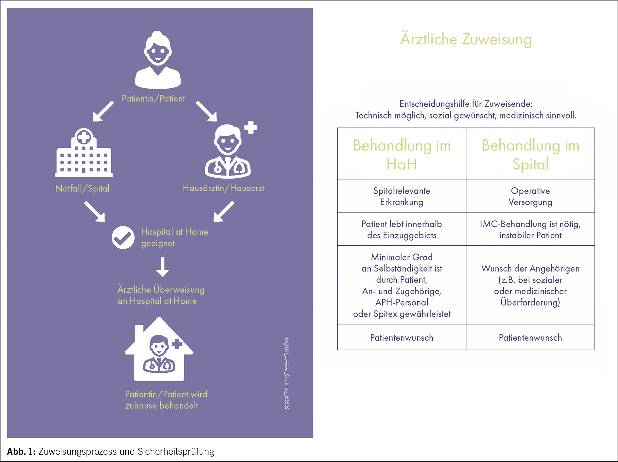
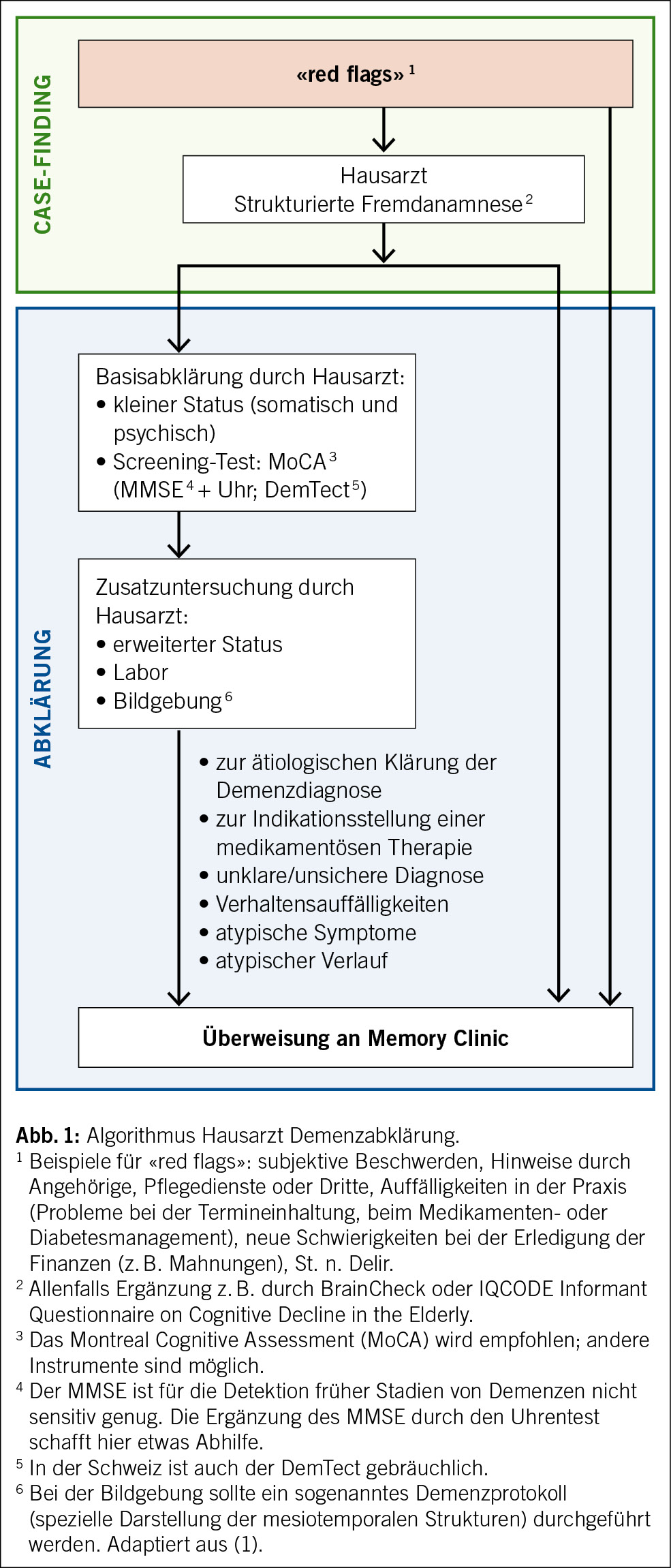
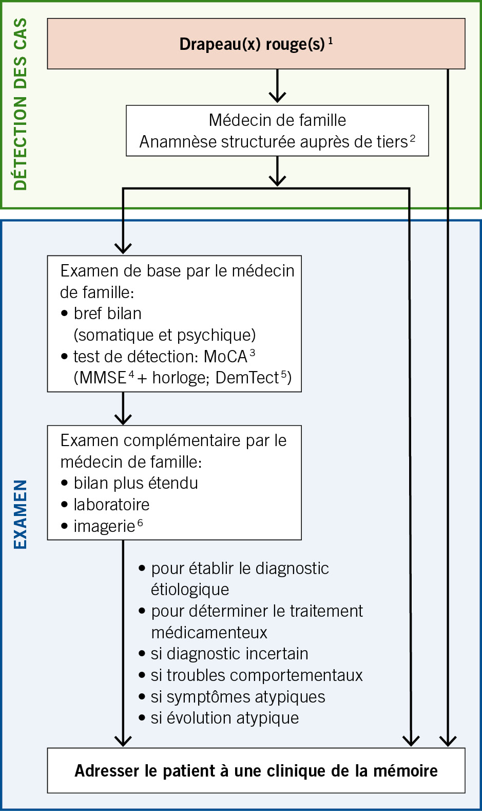
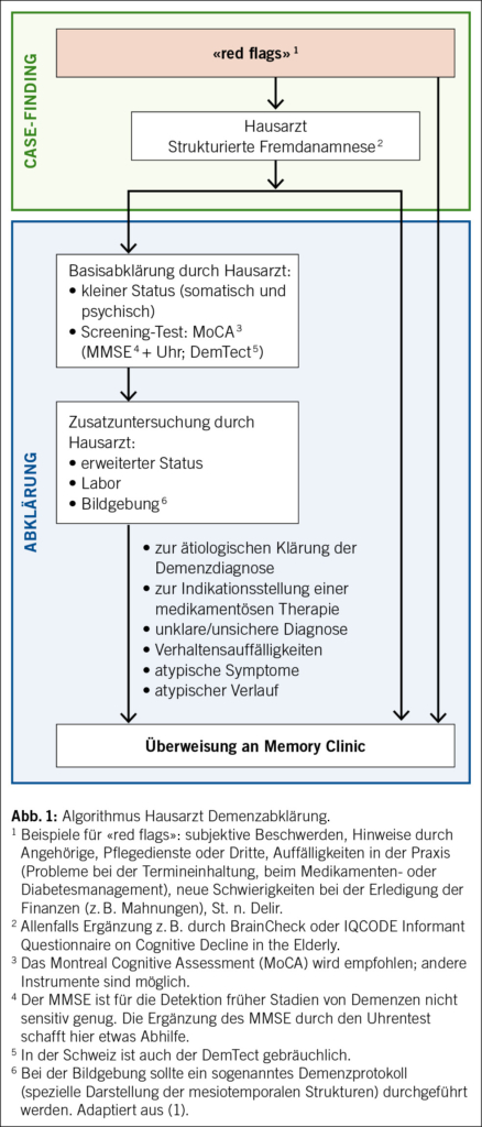
Aufgrund fehlender Evidenz für ein günstiges Kosten-Nutzen-Verhältnis im diagnostischen Prozess (4, 5) wird ein flächendeckendes Screening in höherem Alter nicht empfohlen. Stattdessen sollte ein sogenanntes «case finding» erfolgen. Mit «case finding» ist eine Strategie gemeint, die sich auf Individuen ausrichtet, bei denen Risikofaktoren oder Symptome einer möglichen Demenzerkrankung («red flags») vorhanden sind (4). Das gilt sowohl für Patienten im häuslichen Umfeld als auch für Patienten im Alters- und Pflegeheim. Die Erfassung von «red flags» ist eine Aufgabe der klinischen Grundversorgung (Abb. 1 mit empfohlenem Algorithmus). Je nach Situation kann die anschliessende Abklärung im hausärztlichen Setting oder an einer Memory Clinic erfolgen. Die Abklärung kann bei Bedarf auch anlässlich eines Haus- oder Heimbesuches, z. B. durch geriatrische oder alterspsychiatrische aufsuchende Dienste, erfolgen.
Bei der kognitiven Untersuchung in der Hausarztpraxis ist der Mini-Mental-Status-Examination(MMSE)-Test ein weit gebräuchliches Instrument (6). Die Ergänzung mit dem Uhrentest kann bessere Ergebnisse liefern (7). In der Schweiz ist auch der Demenz-Detektion-Test (DemTect) gebräuchlich (8). Aufgrund der höheren Sensitivität, der kostenlosen Verfügbarkeit in diversen Sprachen sowie der Bewährung im Alltag wird nun das Montreal-Cognitive-Assessment (MoCA) (9) generell für die Anwendung in der Praxis empfohlen.

Ablauf in der Memory Clinic

Interdisziplinäre Diagnostik durch einen Facharzt (Alterspsychiater, Geriater, Neurologe als Mitglieder des Memory-Clinic-Teams oder im Memory-Clinic-Netzwerk verfügbar) und Neuropsychologen:
• Ausführliche Eigen- und Fremdanamnese mittels semistandardisierten Interviews
• Psychiatrischer und somatischer Status
• Neuropsychologische Testung
• Bildgebung (Neuroradiologie, evtl. Nuklearmedizin)
• Laboruntersuchung (Blutentnahme, evtl. Lumbalpunktion)
• Diagnosestellung im Rahmen einer interdisziplinären Diagnosekonferenz mit Diagnosestellung nach ICD-10 oder DSM-5 (Kodierung gemäss ICD-10 oder künftig ICD-11)
• Diagnostische Einordnung der kognitiven Symptome nach Schweregrad sowie nach Präsenz behavioraler und psychischer Symptome der Demenz (BPSD)
• Der Schweregrad der kognitiven Störung wird unter Berücksichtigung der kognitiven Leistungseinbussen und der damit verbundenen Alltagsbeeinträchtigung bestimmt:
– Subjektive kognitive Störungen (kognitive Störung klinisch und neuropsychologisch nicht objektivierbar)
– Leichte kognitive Störung (mild cognitive impairment; minore neurokognitive Störung nach DSM-5)
– Demenz (majore neurokognitive Störung nach DSM-5)
– Leicht: Einschränkungen nur bei IADL (Instrumental Activities of Daily Living) (10), z. B. Haushalt, Umgang mit Geld
– Mittel: Einschränkungen auch bei BADL (Basic Activities of Daily Living) (11), z. B. Nahrungsaufnahme, Ankleiden
– Schwer: vollständige Abhängigkeit
• Stellungnahme zur Ätiologie der kognitiven Störung
• Diagnose- und Beratungsgespräch mit dem Patienten und ihm nahestehenden Personen, siehe unten
• Medikamentöse und nicht medikamentöse Behandlung, siehe Behandlungsempfehlungen der SMC (3)
• Die Memory Clinic ist im Krankheitsverlauf Ansprechpartnerin für verschiedene Fragestellungen wie klinisches Monitoring, Verlaufsdiagnostik und Anpassungen der Behandlungsmassnahmen sowie Durchführung und Vermittlung von Beratung und Begleitung etc.
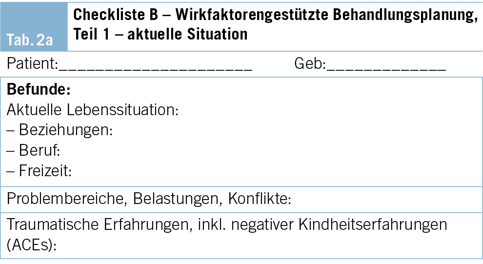
Anamnese

Eine ausführliche Dokumentation und Beschreibung des prodromalen Verlaufs mit der Erfassung von Risikofaktoren, des Krankheitsbeginns und des weiteren Krankheitsverlaufs mit allfälligen Fluktuationen sind essenziell. Eine Familienanamnese mit Sterbealter und Todesursache der Eltern, der Geschwister und allenfalls der Kinder sollte erhoben werden. Falls Demenzerkrankungen in der Familie gehäuft vorkommen, sollten Angaben zum Alter bei Krankheitsbeginn und zum Verlauf festgehalten werden. Zudem wird eine vollständige persönliche Anamnese inklusive Medikamentenanamnese eingeholt.

Systemanamnese

• Noxen: Alkohol, Nikotin, Drogen, Missbrauch von Medikamenten
• Herzkreislauf: kardiopulmonale Beschwerden, Herzrhythmusstörungen (insbesondere Vorhofflimmern)
• Ernährung: Gewichtsverlauf, Nahrungsaufnahme inkl. Zahnstatus/Dysphagie, Verdauung, Defäkationsbeschwerden (z. B. Obstipation), Diäten, Diabetes
• Miktion: Urininkontinenz, Dysurie, häufiges Wasserlassen, Harndrang
• Sensationen: Schwindel, Kopfschmerzen, allgemeine Schmerzen
• Erholung: Tagesschläfrigkeit, Ein- und Durchschlafstörungen, (Alb-)Träume, nächtliche Unruhe, Parasomnien, Schnarchen, Apnoephasen
• Mobilität: Gehhilfen, Stürze (Häufigkeit, Ursache, Ablauf), subjektive Gangunsicherheit
• Neurologische Symptome: Koordinations- und Gleichgewichtsstörungen, Tremor, Tonus, allgemeine Schwäche (motorische und sensible Defizite), Augenmotilitätsstörungen, Dysphagie, Krampfanfälle
• Sprache: undeutliches Sprechen, Wortfindungsschwierigkeiten, Sprachverständnisstörung
• Sinnesorgane: Visus- und Hörverminderung, verändertes Riechen und Schmecken
• Psychische Verfassung: depressive Symptome inkl. Suizidalität, Angst, Reizbarkeit, Apathie, Agitation, Wahn, Sinnestäuschungen
• Deliranamnese
• Am Schluss: noch nicht angesprochene Beschwerden

Psychosoziale Anamnese

Die Demenzdiagnostik wird häufig bei älteren multimorbiden Patienten durchgeführt, bei denen die Kommunikationsfähigkeit und die Krankheitswahrnehmung beeinträchtigt und die Anamneseerhebung erschwert sein können.
In Form eines semistrukturierten Interviews wird mit den Betroffenen und den Angehörigen die eigentliche Problemanamnese zu erlebten und beobachteten Hirnleistungsstörungen im Alltag erhoben. Das erfolgt mit gezielten Fragen zu Gedächtnisleistungen, sprachlichen Fähigkeiten, zum Planungs- und Steuerungsverhalten, zur Kommunikation sowie zeitlichen und räumlichen Orientierungsfähigkeit. Angaben zur Alltagsgestaltung, insbesondere zum Grad der Selbständigkeit in den primären (basalen) und erweiterten (instrumentellen) Alltagsaktivitäten, werden detailliert erfragt. Dazu gehören Informationen zum Unterstützungsbedarf, zu den nötigen Hilfestellungen und bereits aktivierten Helfern. Es werden Informationen zum Autofahren (Fahrsicherheit, Fahrkompetenz) und/oder zur Benutzung öffentlicher Verkehrsmittel eingeholt. Zusätzlich empfiehlt es sich, Angaben zu sozialen Aktivitäten, Hobbys und gepflegten Kontakten zu erheben.
Das Interview wird ergänzt mit präzisen Fragen zur Händigkeit, zur primären und zu weiteren Sprachen sowie zu lebensgeschichtlichen Aspekten wie der Ausbildung und beruflichen Laufbahn, der familiären und aktuellen Lebenssituation. Ergänzt werden die medizinische und die psychosoziale Anamnese in der Regel durch Selbst- und Fremdbeurteilungsskalen, zum Beispiel:
• Informant Questionnaire on Cognitive Decline in
the ­Elderly (IQCODE)
• Nurses’ Observation Scale for Geriatric Patients (NOSGER)
• Caregiver Burden Inventory (CBI)
• Neuropsychiatrisches Inventar (NPI)
• Geriatrische Depressionsskala (GDS)
• Epworth Sleepiness Scale (ESS)

Erhebung der Alltagsfähigkeiten

Bei den Alltagsfähigkeiten (Activities of Daily Living, ADL) wird zwischen basalen (BADL) und instrumentellen Fähigkeiten (IADL) unterschieden. Die BADL umfassen die Bereiche Essen, Nahrungsaufnahme, Körperpflege, Ankleiden, Toilettengang, Kontinenz, die Fähigkeit, aus dem Bett oder von einem Stuhl aufzustehen, sowie Fortbewegung und Mobilität (mit oder ohne Gehhilfe). Zu den IADL werden folgende Kompetenzen gezählt: Mahlzeiten zubereiten, den Haushalt und die Wäsche erledigen, finanzielle Angelegenheiten erledigen, telefonieren, einkaufen, Medikamenteneinnahme, Fortbewegung unter Nutzung eines Transportmittels.
Die Erhebung der Alltagsfähigkeiten eines Patienten stützt sich im Rahmen der Anamnese und Fremdanamnese grundsätzlich auf vier mögliche Quellen:
1. Beobachtung des Patienten
2. Standardisierte Befragung des Patienten
3. Standardisierte Befragung der Angehörigen oder ­Betreuungspersonen

Erhebung bei aufsuchender Abklärung (Hausbesuch, im Heim etc.)

Dabei ist immer zwischen früher vorhandenen und aktuellen Fähigkeiten zu unterscheiden und besonders auf Diskrepanzen zwischen Eigen- und Fremdanamnese zu achten. Aufschlussreich kann auch die Frage sein, ob sich jemand neuerer Technologien (IADL) bedient oder lieber beim Altbewährten bleibt. Die strukturierte Erfassung der Alltagsfähigkeiten im Rahmen einer persönlichen, bei Bedarf vom Patienteninterview getrennt durchgeführten Fremdanamnese (12) ist zwingender Bestandteil jeder Demenzabklärung.
Eine aufsuchende Abklärung der ADL ist im Rahmen der Untersuchungen in der Memory Clinic meist aus praktischen Gründen nicht möglich. Diese kann z. B. durch aufsuchende Dienste oder Fachpersonen in einem separaten Schritt erfolgen.

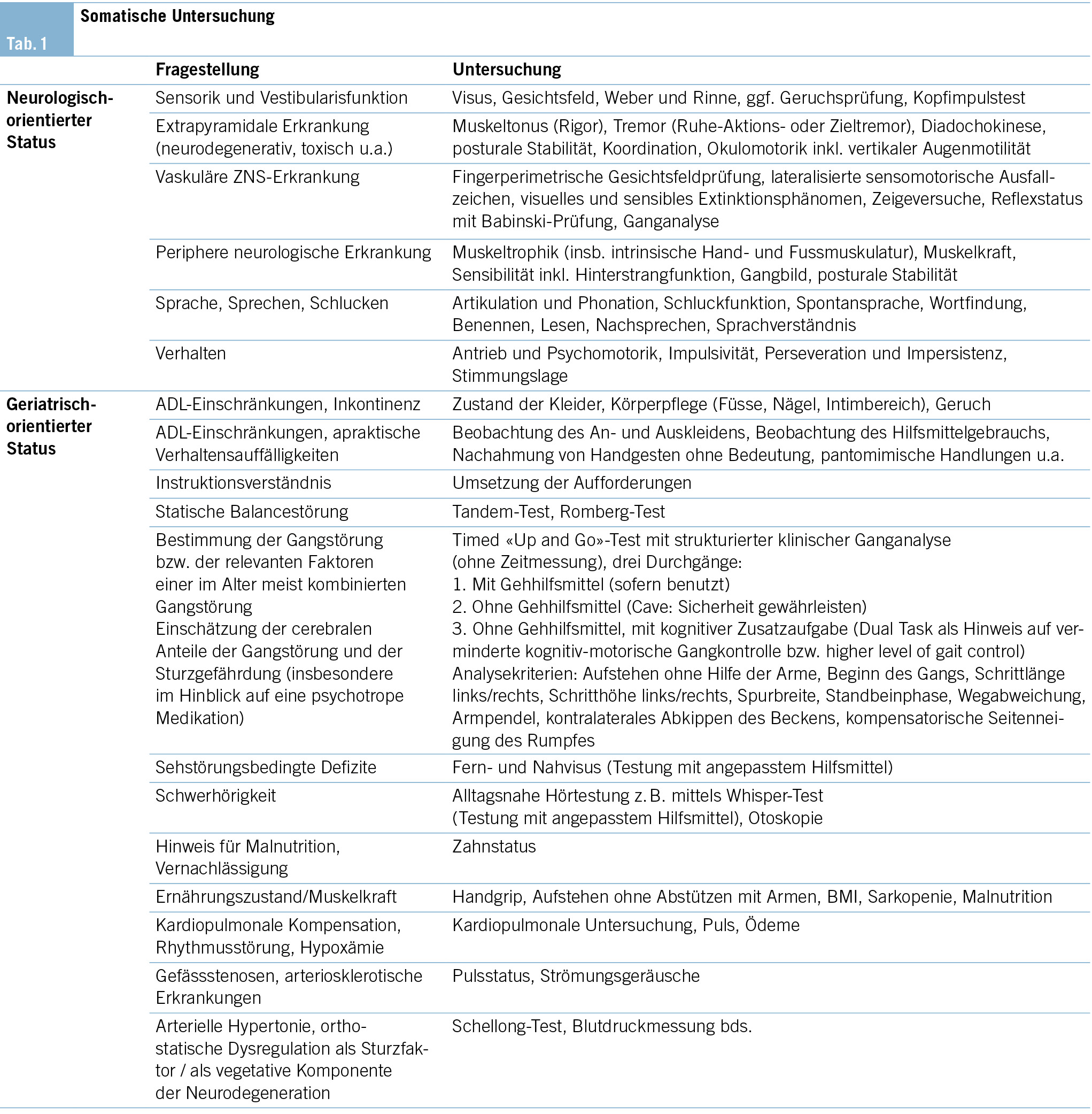
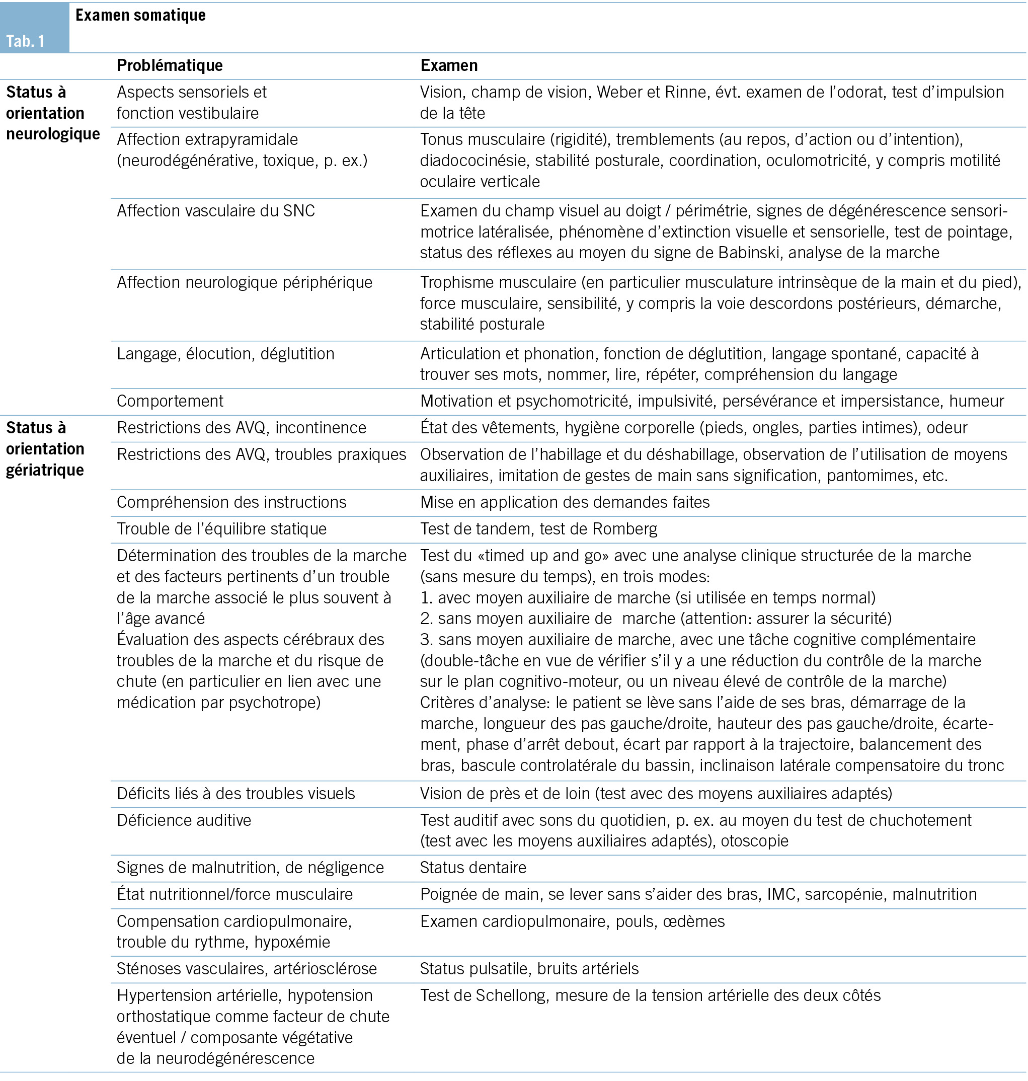
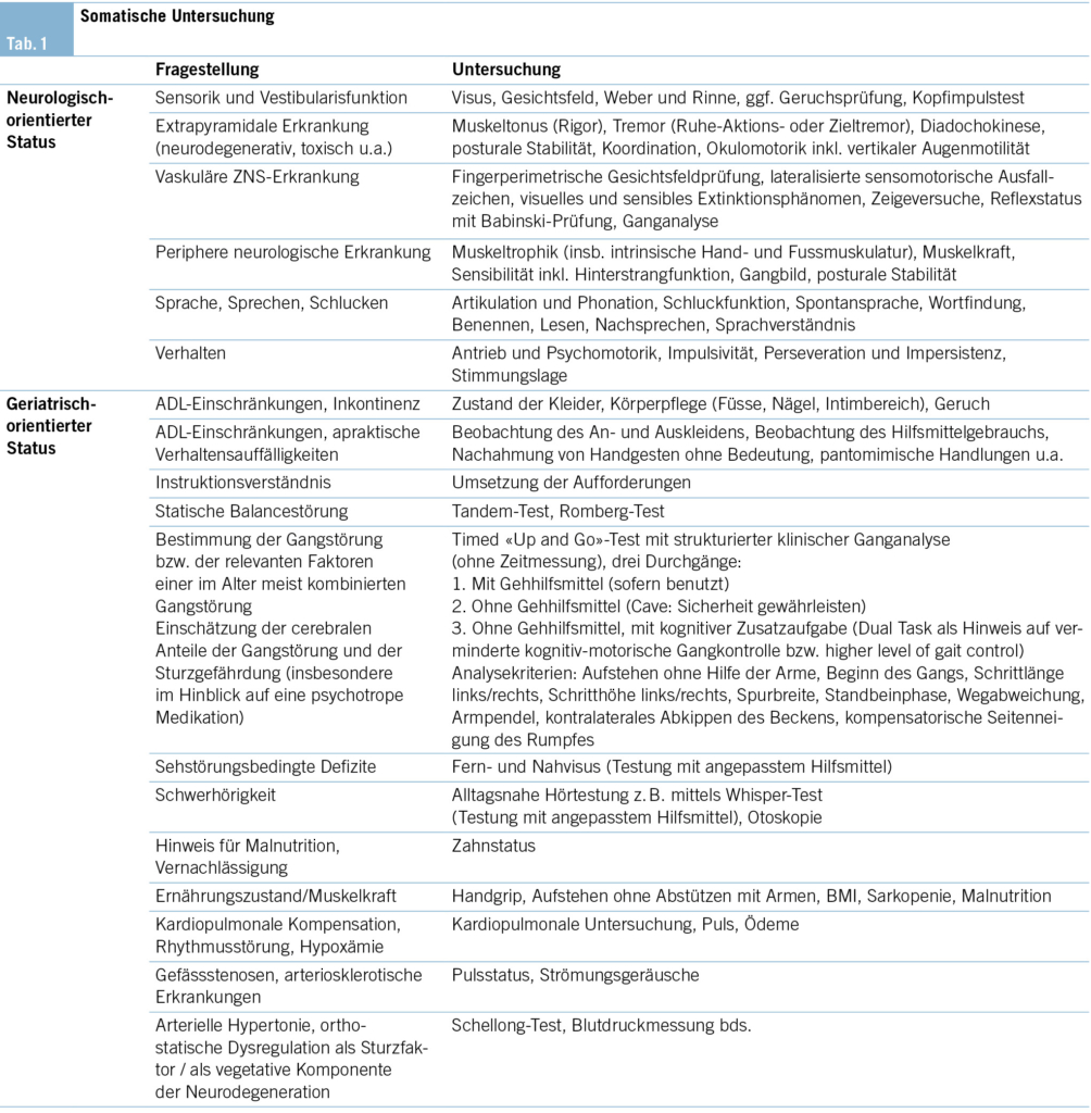
Somatische Untersuchung

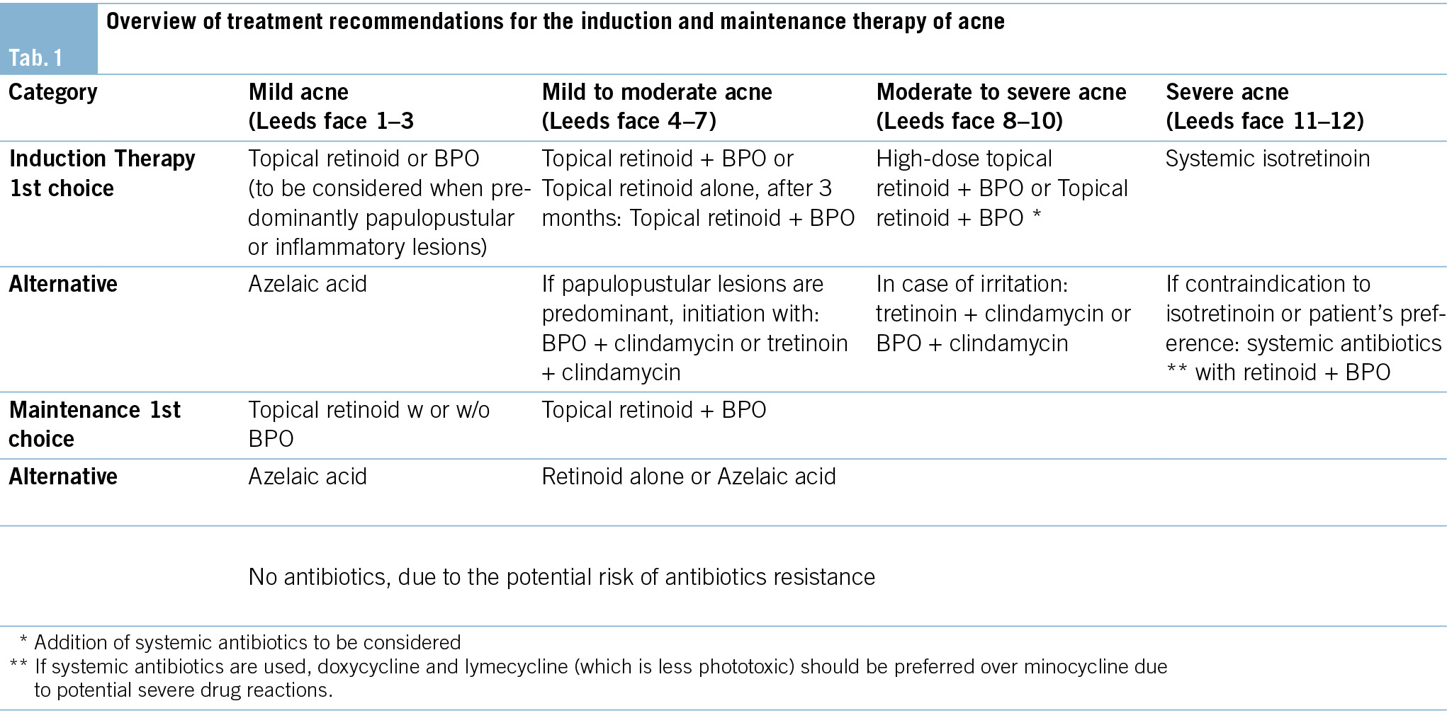
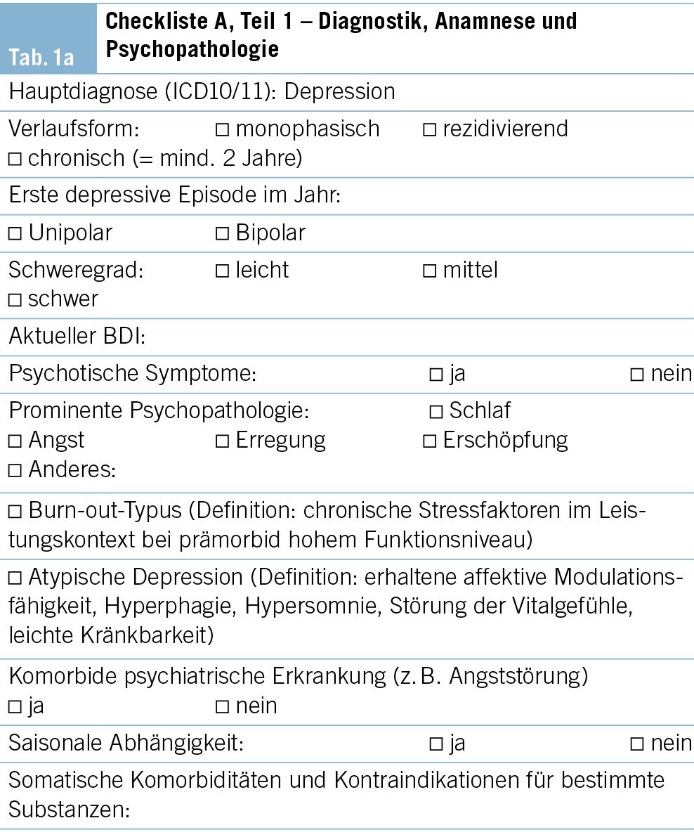
Die somatische Untersuchung richtet sich nach den Komorbiditäten und der Verdachtsdiagnose (Schwerpunkt der Fragestellung) und muss darauf entsprechend angepasst werden (Tab. 1).

Psychopathologischer Status und ­behaviorale und psychische Symptome der Demenz (BPSD)

Nebst kognitiven Störungen und Einschränkungen bei Alltagsaktivitäten treten im Verlauf der Demenzerkrankungen oft verschiedene psychiatrische und Verhaltenssymptome auf, welche die Betroffenen erheblich beeinträchtigen und ihre Behandlung und Pflege erschweren können (13). Diese Symptome können auftreten als Ausdruck von psychischen Störungen, welche unabhängig von der Demenzerkrankung vorbestehen, oder als Symptome der Demenzerkrankung. Für neuropsychiatrische Symptome, die im Rahmen oder als Folge der Demenzerkrankung auftreten, wurde die Bezeichnung behaviorale und psychische Symp­tome der Demenz (BPSD) vorgeschlagen. Wesentlich für die Beurteilung und die Behandlung ist, sowohl biologische als auch psychologische und Umweltfaktoren zu berücksichtigen. Zu erwähnen ist, dass neuropsychiatrische Symp­tome nicht erst im Stadium der Demenz, sondern bereits bei MCI auftreten und sogar den kognitiven Störungen vorausgehen können (14, 15). Früh auftretende neuropsychiatrische Symptome sind mit einer schnelleren kognitiven und alltagsfunktionalen Verschlechterung assoziiert (16).

Standard

• Klinische Untersuchung auf das Vorliegen psychischer Symptome:
– Subjektive Bewertung durch den Patienten
– Fremdbewertung durch Angehörige oder Personen, die den Patienten gut kennen
– Psychopathologischer Grundstatus
– Beurteilung hinsichtlich des Vorliegens einer psychia­trischen Begleiterkrankung, insbesondere einer depressiven Störung oder eines Delirs
• Gewichtung der Informationen im Diagnoseprozess
• Berücksichtigung primärer psychiatrischer Begleiterkrankungen und neuropsychiatrischer Symptome/BPSD bei Therapievorschlägen zu kognitiven Störungen
• Überweisung an spezialisierte Sprechstunde, insbesondere bei mittelgradig bis schwer ausgeprägten psychiatrischen Symptomen, bei bestätigten oder vermuteten psychiatrischen Begleiterkrankungen, bei komplexen Implikationen für die Familie oder bei potenziell erforderlichen freiheitsbeschränkenden Massnahmen

Optional

• Die Anwendung eines psychopathologischen Beschreibungsrasters wird empfohlen (AMDP).
• Der Einsatz spezieller psychopathologischer Skalen wird empfohlen, z. B. das Neuropsychiatric Inventory Questionnaire (17); bei V. a. Depression: Geriatrische Depressionsskala (18) oder Beck Depression Inventory (19) – wenn sich der Patient noch im Frühstadium der Demenzerkrankung befindet.
• Die Behandlung der psychischen Begleiterkrankungen an der Memory Clinic: je nach Kompetenz und Verfügbarkeit.

Neuropsychologische Untersuchung

Der wichtigste Beitrag der kognitiven Testung im Rahmen einer neuropsychologischen Abklärung sind die Früherkennung und Quantifizierung der beeinträchtigten kognitiven Dimensionen. Zudem liefert ein kognitives Stärken-Schwächen-Profil eine wichtige Grundlage für die individuelle Psychoedukation, Beratung und Therapie der Patienten und deren Angehörigen. In Anlehnung an die für neurokognitive Störungen differenzierte Darstellung im DSM-5 (20) muss die umfassende neuropsychologische Untersuchung qualitative und quantitative Aussagen zu folgenden sechs Dimensionen liefern:
• Komplexe Aufmerksamkeit: Daueraufmerksamkeit, geteilte Aufmerksamkeit, selektive Aufmerksamkeit, Verarbeitungsgeschwindigkeit
• Exekutive Funktionen: planen, entscheiden, Arbeitsgedächtnis, verwerten von Feedback/Fehlerkorrektur, handeln entgegen der Gewohnheit/Verhaltenshemmung, mentale Flexibilität
• Lernen und Gedächtnis: unmittelbares Gedächtnis, Kurzzeitgedächtnis (inkl. freier Abruf, Abruf mit Hinweisreizen, Wiedererkennen), Langzeitgedächtnis (semantisch, autobiografisch), implizites Lernen
• Sprache: Sprachproduktion (inkl. Benennen, Wortfindung, Wortflüssigkeit, Grammatik, Syntax), Sprachverständnis
• Perzeptiv-motorische Fähigkeiten: visuelle Wahrnehmung, Visuokonstruktion, perzeptuell-motorische Fähigkeiten, Praxis, Gnosis
• Soziale Kognition: Emotionen erkennen, Empathiefähigkeit, « theory of mind », Verhaltensänderungen
Die Beurteilung der Leistungen soll beruhen auf:
• der gemessenen Leistung (mindestens zwei Testverfahren pro Domäne, z. B. verbale und visuelle Gedächtnisleistung) in den Tests wie auch auf
• der Beobachtung/Befragung.
Mit Ausnahme der Dimension «soziale Kognition» (hierfür sind Instrumente derzeit noch in Entwicklung) existieren für alle Aspekte zeitlich ökonomisch einsetzbare kognitive Testinstrumente. Es sind Instrumente zu verwenden, die über eine Normierung verfügen, welche Alter, Geschlecht und Ausbildung der untersuchten Personen berücksichtigen.

Praktisches Vorgehen

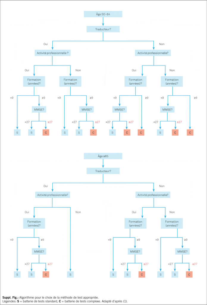
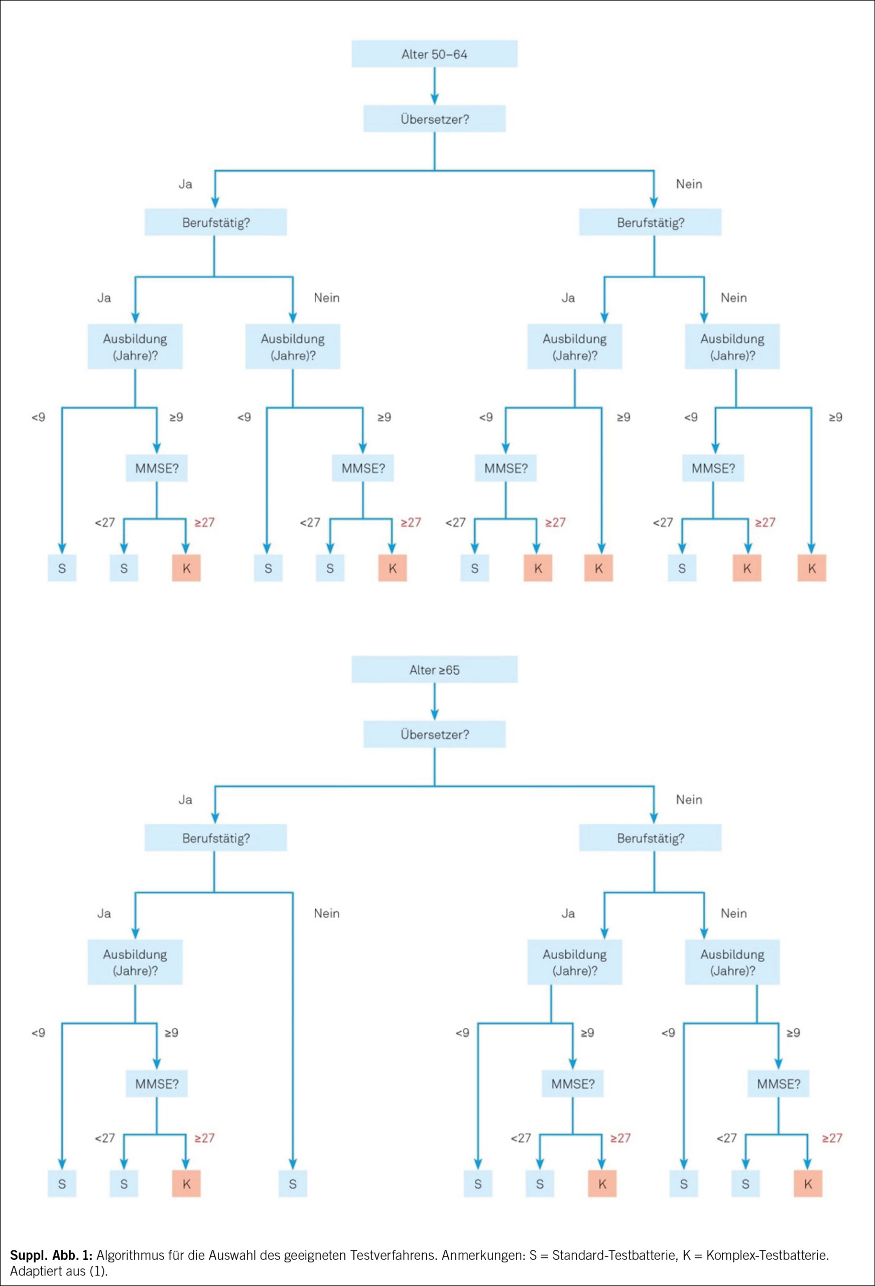
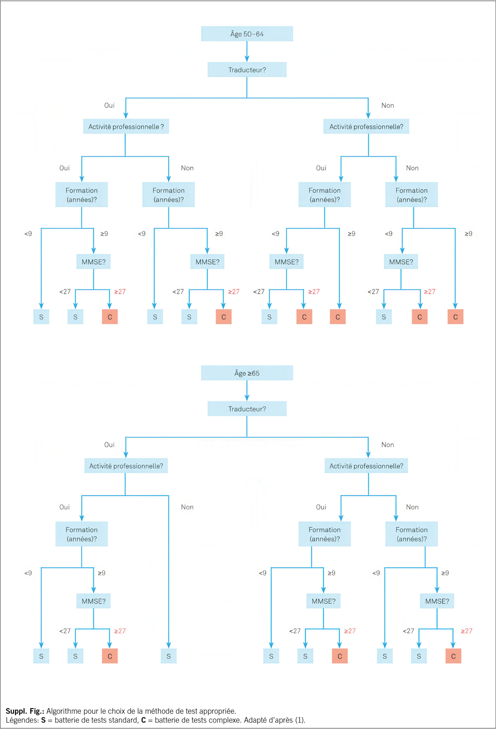
Zum etablierten Standard gehört die Durchführung eines kognitiven Kurztests, dessen Resultate unter Berücksichtigung der Informationen aus Anamnese, Fremdanamnese erlauben, die richtige Testauswahl für die umfassende hypothesengeleitete und personalisierte neuropsychologische Untersuchung zu treffen. MoCA (9) oder MMSE (6), kombiniert mit dem Uhrentest (7), wird als fester Bestandteil für das standardisierte Kurzverfahren empfohlen. Für Patienten mit fortgeschrittener Demenz kann sich eine differenzierte kognitive Testung erübrigen. Bei der Auswahl der geeigneten umfassenden kognitiven Testbatterie kann ein Algorithmus verwendet werden (Suppl. Abb. 1). Bei kognitiv nicht stark beeinträchtigten Patienten in einer Memory Clinic gehört die umfassendere neuropsychologische Untersuchung zum Standard. Eine in der deutschen Schweiz sehr gebräuchliche, bestens normierte und validierte Testbatterie ist die «Consortium to Establish a Registry for Alzheimer’s Disease – Neuropsychological Assessment Battery (CERAD-NAB)», ergänzt mit Tests zur Untersuchung der Verarbeitungsgeschwindigkeit und Exekutivfunktionen, die als «CERAD-Plus» bezeichnet wird. Die Testunterlagen der CERAD-Plus inkl. der Möglichkeit zur Auswertung stehen Fachpersonen unter www.memoryclinic.ch kostenlos zur Verfügung. Eine ausführlichere Liste von Tests findet sich unter www.swissmemoryclinics.ch.
Eine solche Standarduntersuchung wird durch weitere neuropsychologische Testinstrumente ergänzt, wenn ein hohes kognitives Ausgangsniveau besteht, sich Fragen zum Beispiel zur Arbeitsfähigkeit stellen sowie bei seltenen Demenzformen mit spezifischen kognitiven Ausfallmustern.

Untersuchung spezieller Patientengruppen

Eine besondere Herausforderung stellen Patienten dar, die: (a) einen Migrationshintergrund mit eingeschränkter Sprachkompetenz in der Untersuchungssprache haben, (b) sensorische Einschränkungen haben, für welche in der Regel keine Normwerte existieren, (c) intellektuelle Entwicklungsstörungen haben, (d) Hochbegabung aufweisen oder (e) hochbetagt sind. Hier müssen die kognitive Untersuchung und Beurteilung auf die individuelle Situation angepasst werden, und es erfordert eine umfassende neuropsychologische Expertise. Darüber hinaus können somatische und psychiatrische Erkrankungen sowie eine die Kognition beeinträchtigende Medikation die Beurteilung erschweren und müssen in jedem Fall berücksichtigt werden.

Beurteilung und Interpretation

Die empfohlene Einteilung zum Schweregrad ist die sieben-kategorielle Klassifikation der «Leitlinien zur Klassifikation und Interpretation neuropsychologischer Testergebnisse» der Schweizerischen Vereinigung für Neuropsychologie (SVNP): https://neuro.psychologie.ch. Im Bericht über die neuropsychologische Untersuchung soll transparent sein, auf welche Art von Daten sich die Schlussfolgerungen beziehen. Zudem muss eine kurze Stellungnahme zum klinischen Eindruck erfolgen. Es ist sehr hilfreich, die Resultate der kognitiven Testung grafisch darzustellen. Es ist dann sinnvoll und erwünscht – gestützt auf die Resultate der kognitiven Testung und auf funktionell-neuroanatomischen Überlegungen –, Hypothesen zur Ätiologie einer vorhandenen Störung aufzustellen.

Labordiagnostik

Blutdiagnostik

Standard
• Blutbild, C-reaktives Protein
• Glukose
• Natrium, Kalium, Kalzium korrigiert
• Kreatinin, eGFR
• GOT (Glutamat-Oxalacetat-Transaminase), GPT (Glu­tamat-Pyruvat-Transaminase), γ-GT (Gamma-Glutamyl-Transferase)
• TSH (Thyreoidea-stimulierendes Hormon)
• Vitamin B12, Folsäure
• Cholesterin, HDL-Cholesterin, Triglyceride (Lipidstatus bei unter 80-Jährigen)

Ergänzende Untersuchungen bei spezifischem Verdacht oder pathologischen Resultaten der Standarddiagnostik:
• Vitamin D
• Lues- und Borrelienserologien, HIV-Test
• CDT (Carbohydrat-defizientes Transferrin)
• fT3, fT4, Parathormon, Kortisol
• Differenzialblutbild, BSG, INR
• CK (Creatinkinase), Harnstoff, Harnsäure, Bilirubin, Phosphat, Chlorid, Magnesium, Zink
• Blutzuckertagesprofil, HbA1c
• Vitamine B1 und B6, Niacin, Homocystein, Holotranscobolamin und/oder Methylmalonsäure
• Ferritin, Transferrin
• Kupfer, Coeruloplasmin, Urinstatus mit Kupfer-Clearance im 24-Stunden-Urin
• Noxenscreening (Blei, Quecksilber)
• Drogenscreening (z. B. Benzodiazepine)
• Drug-Monitoring
• Autoimmune und paraneoplastische Encephalitis-Antikörper
• Vaskulitisparameter
• APOE-Genotyp (z. B. im Rahmen der Forschung, bei geplanter Anti-Amyloid-Therapie mit monoklonalen Antikörpern)

Liquordiagnostik

Standard für folgende Indikationen (21): Ausschluss nicht primär degenerativer Demenzformen, hier insbesondere chronisch-entzündlicher ZNS-Erkrankungen:
• Bei rapid-progredienten, atypischen oder frühen (Erstmanifestation vor dem 65. Lebensjahr) Demenzerkrankungen
• Diagnostische- und Entlastungspunktion bei Verdacht auf Normaldruckhydrozephalus
• Unterstützende Diagnostik für den Nachweis von Neurodegeneration, Tau-Pathologie und/oder Amyloid-Pathologie; bei spezifisch gestellter Indikation und Aufklärung bei Verdacht auf Frühstadien der Alzheimer-Krankheit (einschliesslich des Stadiums der leichten kognitiven Störung, MCI)
Neben der Basisdiagnostik Liquor sollte bei Verdacht auf eine chronisch-entzündliche ZNS-Erkrankung eine zusätzliche Bestimmung der oligoklonalen Banden sowie gegebenenfalls Borrelien- und Lues-Serologien und weitere serologische Bestimmungen durchgeführt werden. Die Bestimmung von Beta-Amyloid (Aβ42, Aβ42/Aβ40), phosphoryliertes Tau (pTau) und Gesamt-Tau als Demenzbiomarker eignet sich als früh- und differenzialdiagnostisches Verfahren bei der Abklärung kognitiver Störungen und wird meist als Teil der Basisdiagnostik Liquor durchgeführt. Die obligatorische Krankenpflegeversicherung (OKP) übernimmt unter bestimmten Limitationen die Kosten für Analysen von Liquormarkern, auch bei Patienten mit MCI. Bei klinischem Verdacht auf Creutzfeldt-Jakob-Erkrankung können eine zusätzliche Bestimmung des Proteins 14-3-3 und eine RT-QuIC (Real-Time Quaking-Induced Conversion) im Liquor erfolgen. Eine individuell angepasste Aufklärung der Patienten zur geplanten Biomarkerdiagnostik und den möglichen Ergebnissen wird empfohlen (21).

Bildgebende Diagnostik

Neuroradiologische Verfahren

Die bildgebende Untersuchung des Gehirns im Rahmen der Diagnostik von Demenzerkrankungen erfüllt zwei wesentliche Funktionen:
1. Ausschluss von sekundären Demenzursachen, wie beispielsweise Raumforderungen, Liquorzirkulationsstörungen sowie vaskulär, metabolisch oder entzündlich bedingten Veränderungen
2. Beitrag zur ätiologischen Differenzierung und Zuordnung primärer Demenzerkrankungen
Die Bildgebung des Gehirns kann zur Diagnosestellung und Differenzialdiagnose zwischen der Alzheimer-Erkrankung und anderen, z. B. vaskulären oder frontotemporalen, Demenzen beitragen, wobei die differenzialdiagnostische Trennschärfe der strukturellen Bildgebung zwischen diesen Erkrankungen derzeit für die alleinige Anwendung nicht ausreichend ist (22). Ein wesentlicher Nutzen der strukturellen Bildgebung des Gehirns besteht in der Identifizierung und Beurteilung intrazerebraler Läsionen bezüglich Lokalisation und Quantität. Die Bildgebung ist dabei als ein Beitrag zur Gesamtbeurteilung und differenzialdiagnostischer Einordnung der demenziellen Krankheitsbilder in Verbindung mit Anamnese und den klinischen und neuropsychologischen und sonstigen Befunden anzusehen.

Standard
Die strukturelle Bildgebung mit kranialer Magnetresonanztomografie (MRT) – alternativ, bei Vorliegen von MRT-Kontraindikationen mittels kranialer Computertomografie (CT) – ist Teil der Standarddiagnostik.

Kraniale Magnetresonanztomografie (MRT)
Aufgrund der fehlenden Strahlenbelastung und des wesentlich besseren Gewebekontrastes im Gehirn im Vergleich zur CT sollte die MRT als Untersuchungsmethode der Wahl eingesetzt werden. Falls vorhanden, sollte ein 3- Tesla-MRT einem 1.5-Tesla-MRT vorgezogen werden. Die detaillierten Empfehlungen zum Untersuchungsprotokoll sind auf der SMC-Website zu finden (www.swissmemoryclinics.ch).

Kraniale Computertomografie (CT)
Bei fehlender Verfügbarkeit der MRT und bei Kontra­indikationen für eine MRT (z. B. Herzschrittmacher, digital programmierte Implantate, ausgeprägte Klaustrophobie) sollte alternativ eine CT durchgeführt werden. Die CT ohne Kontrastmittel ist in der Regel ausreichend für den Nachweis oder Ausschluss von Raumforderungen, eines subduralen Hämatoms oder eines Hydrozephalus, mit Einschränkung auch für die Abklärung einer vaskulären Demenz.

Sonografie der hirnversorgenden Gefässe
Bei vaskulärer Demenz oder bei gemischt vaskulär-degenerativen Demenzformen kann die Beurteilung von Stenosen der hirnversorgenden Gefässe mittels Doppler- und Duplexsonografie relevant sein.

Nuklearmedizinische Verfahren

Positronen-Emissions-Tomografie (PET) mit 18F-Fluordesoxyglukose (FDG-PET)
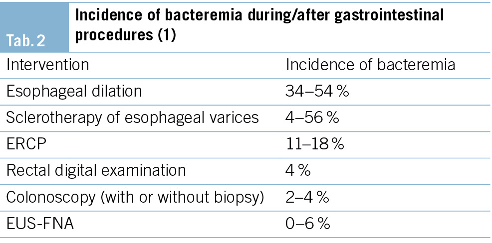
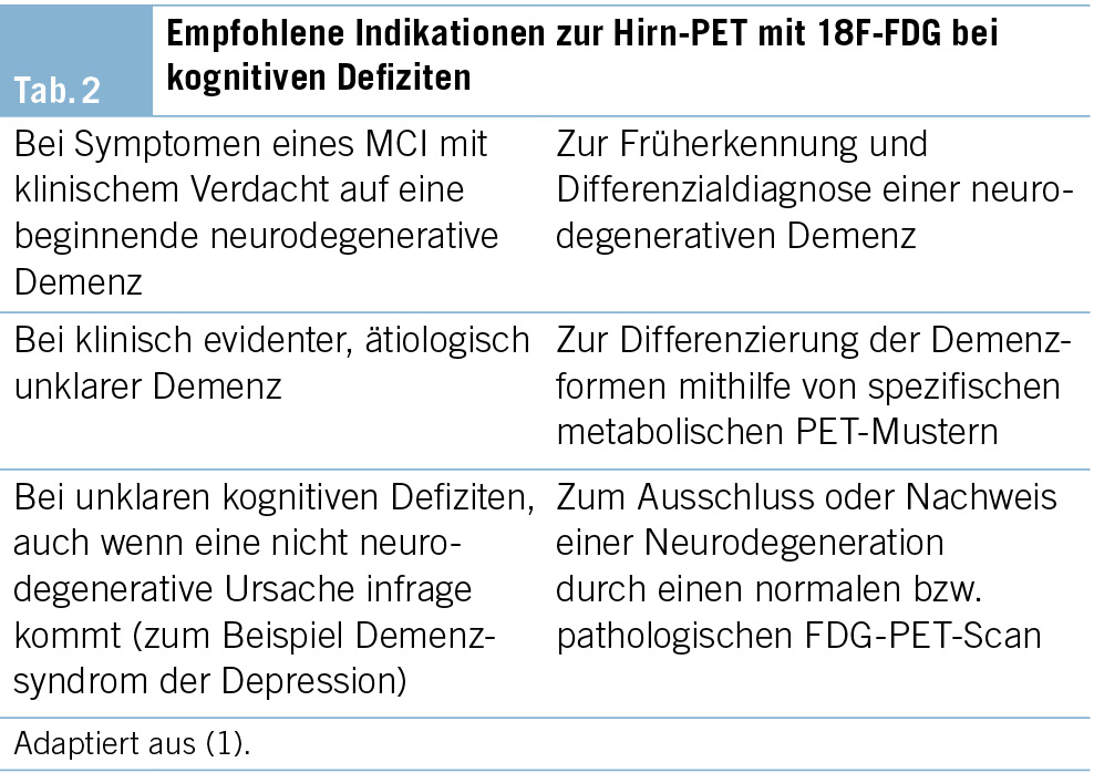
Die FDG-PET ermöglicht die bildliche Darstellung des regionalen Glukosemetabolismus des Gehirns und den Vergleich mit Normalwerten. Die FDG-PET ist die am besten validierte nuklearmedizinische Bildgebung zur Abklärung von Demenzen. Die empfohlenen Indikationen zur FDG-PET sind in der Tab. 2 aufgeführt.
Die FDG-PET weist eine hohe Sensitivität für den Nachweis funktioneller und neurodegenerativer Veränderungen der kortikalen Informationsverarbeitung auf und eignet sich hervorragend zur Frühdiagnostik, auch im Stadium einer leichten kognitiven Störung (MCI) (23). Die topische Zuordnung funktionsgestörter Areale ermöglicht zudem häufig die Differenzierung zwischen früher Neurodegeneration, funktionellen, demenzähnlichen Zuständen (z. B. bei psychiatrischen Erkrankungen) und kognitiven Beeinträchtigungen anderer Genese (z. B. limbische Enzephalitis).
Auch für eine Reihe weiterer, seltenerer neurodegenerativer Demenzen bestehen typische metabolische Muster, welche mittels der FDG-PET differenziert werden können. Dies betrifft die Demenz mit Lewy-Körperchen, verschiedene Formen der frontotemporalen Lobärdegenerationen, hier insbesondere die Varianten der primär progressiven Aphasie (semantische, nicht flüssige und logopenische), sowie die posteriore kortikale Atrophie (24).
Zusammenfassend wird die FDG-PET für die Diagnostik neurodegenerativer Demenzen als molekulare Bildgebungsmethode der ersten Wahl empfohlen (23). Für die Kostenübernahme gemäss Krankenpflege-Leistungsverordnung (KLV) sind Limitationen zu beachten.

Amyloid-PET
Die Amyloid-PET kann zuverlässig nachweisen, ob die Beta-Amyloid-Pathologie im Gehirn vorliegt oder nicht. Dabei können verschiedene Tracer eingesetzt werden, die an Amyloid-Plaques binden. Seit 01.04.2020 besteht eine Leistungspflicht der Krankenkassen für Amyloid-PET (Limitationen sind zu beachten) als weiterführende Untersuchung in unklaren Fällen nach inkonklusiver Liquordiagnostik oder wenn eine Lumbalpunktion nicht möglich oder kontraindiziert ist (23).
Bei künftig erwarteter Verfügbarkeit neuer Arzneimittel, die sich gegen Amyloid-Pathologie richten, zeichnet sich ein erweitertes klinisches Einsatzgebiet ab.

Bildgebung des dopaminergen Systems: Dopamin-Transporter-SPECT mit 123I-Ioflupane und 18F-DOPA-PET
Bei der Dopamin-Transporter-SPECT mit 123I-Ioflupane und der PET mit 18Fluoro-DOPA handelt es sich um eine nuklearmedizinische Untersuchung, um die zerebrale Dopamin-Verfügbarkeit zu bestimmen (25). Der Nachweis eines striatalen dopaminergen Defizits durch diese Verfahren kann zuverlässig zwischen einer DLB (mit pathologischem Befund im Striatum) und Demenzen ohne Lewy-Körperchen unterscheiden (26). Für zwei SPECT-Radiopharmaka (DaTSCAN® und Striascan®) sowie für 18F-DOPA-PET wurde die Swissmedic-Zulassung für diese Indikation erteilt.

Genetische Untersuchung

Eine häufig von Betroffenen von Demenzerkrankungen und ihren Angehörigen gestellte Frage ist, ob die Erkrankungen, die sie betreffen, vererbt werden. Die Mitarbeiter einer Memory Clinic müssen in der Lage sein,
• Fälle der familiär auftretenden Demenzerkrankungen zu identifizieren;
• Informationen im Rahmen der Diagnosestellung zu gewichten;
• die Indikation für eine genetische Beratung zu stellen;
• Aufklärung und Beratung zu genetischen Risikofaktoren für die häufigsten Demenzerkrankungen zu leisten;
• den Patienten bzw. dessen Familie an ein humangenetisches Institut zu verweisen (meist an eine Universitätsklinik angeschlossen). Überweisungen sind insbesondere in folgenden Fällen indiziert:
– ein Angehöriger ersten Grades mit möglicher Erkrankung, Alter unter 50 Jahre
– zwei Angehörige ersten Grades mit möglicher Erkrankung, Alter unter 60 Jahre

Weitere Untersuchungsverfahren

Basierend auf Anamnese oder auf Befunden im somatischen Status kommen weitere Untersuchungen als Ergänzung in Betracht.

Elektroenzephalografie (EEG)

Eine EEG-Untersuchung kann nützliche Informationen liefern bei:
• erheblichen Schwankungen von Vigilanz und Orientierung zum Ausschluss einer epileptischen Genese
• bei V. a. entzündlich-infektiöse, entzündlich-autoimmune oder metabolische ZNS-Erkrankungen (e. g. limbische Encephalitis, Hashimoto-Encephalopathie) sowie bei V. a. Creutzfeldt-Jakob-Erkrankung

Schlafdiagnostik

Demenzerkrankungen gehen häufig mit einem erhöhten Schlafbedürfnis, d. h. einem verlängerten Nachtschlaf und einer erhöhten Tagesschläfrigkeit, einher. Schlafassoziierte Atemstörungen können, ebenso wie hirnorganische Erkrankungen, die Schlafarchitektur stören und eine ­exzessive Tagesschläfrigkeit sowie Einschränkungen von Aufmerksamkeitsleistungen verursachen. Sie stellen einen unabhängigen Risikofaktor für kardio- und zerebrovaskuläre Ereignisse dar und sind häufig behandelbar.
Eine exzessive Tagesschläfrigkeit kann einfach mit dem Epworth-Sleepiness-Fragebogen (27) abgeschätzt werden. Die Durchführung einer apparativen schlafbegleitenden Untersuchung (Aktimetrie, nächtliche Pulsoximetrie, Polygrafie, Polysomnografie) kann indiziert sein bei:
• einer exzessiven Tagesschläfrigkeit und Zeichen einer nächtlichen Atemstörung zum Ausschluss eines Schlaf-Apnoe-Syndroms
• V.a. REM-Schlaf-Verhaltensauffälligkeiten
• V.a. schlafgebundene epileptische Anfälle

Geruchstestung

Eine Geruchstestung kann bei der Frühdiagnostik der Verdachtsdiagnose Alzheimer-Demenz unterstützende Befunde liefern. Bei der Alzheimer- sowie bei der Parkinson-Krankheit kommt es frühzeitig zu einer Beeinträchtigung der Geruchsleistung. Es stehen verschiedene validierte Tests zur Verfügung.

Ganganalyse

Neurodegenerative Erkrankungen können neben der Ko­gnition auch motorische Funktionen beeinträchtigen. Menschen mit einer demenziellen Erkrankung haben gegenüber gleichaltrig Gesunden früh ein erhöhtes Sturzrisiko. Eine strukturierte klinische Ganganalyse ermöglicht hier eine Bewertung der Gangsicherheit bzw. der Sturzgefährdung. Sie hilft, die Indikation für geeignete therapeutische und präventive Massnahmen (Training, Gehhilfsmittel u. a.) zu stellen. Für eine quantitativ ausgerichtete Ganganalyse stehen validierte klinische Mobilitätstests (e. g. Timed «Up and Go»-Test), aber auch computerisierte Verfahren (Bewegungsdaten werden digital erfasst, z. B. über Drucksensoren auf einem Laufteppich) zur Verfügung.

Okulomotorik-/Gesichtsfelduntersuchung

Der klinischen Untersuchung der Okulomotorik und des Gesichtsfeldes kommt bei V. a. bestimmte neurodegenerative Erkrankungen (z. B. eine progressive supranukleäre Blickparese) oder einer vaskulären Pathologie eine besondere Bedeutung zu.

Das Diagnosegespräch

Das Diagnosegespräch folgt der interdisziplinären Dia­gnosestellung und ist der Ausgangspunkt für die individuell angepasste Beratung, Behandlung und Begleitung der Betroffenen. Generell haben Patienten das Recht, klar und angemessen über ihren Gesundheitszustand informiert zu werden, ausser sie verzichten explizit darauf, aufgeklärt zu werden (28). Aufgrund der Tragweite der Diagnose sollte die Übermittlung mit grosser Sorgfalt erfolgen. Einfache, klare, gegebenenfalls den kognitiven Einschränkungen angepasste Worte erleichtern das Verständnis der Diagnose und deren Bedeutung und ermöglichen in der Folge eine offene Kommunikation innerhalb der Familie oder des Bezugssystems. Den Patienten und ihren Angehörigen muss ausreichend die Möglichkeit angeboten werden, Fragen zu stellen (29). Bei Bedarf sollte ein weiteres ergänzendes Beratungsgespräch angeboten werden. Das Wissen über die Erkrankung, deren Verlauf, über mögliche künftige Probleme (z. B. Verhaltensauffälligkeiten, Pflegebedarf) und den Umgang mit diesen hilft, die emotionale Belastung zu verringern. Über Unterstützungs- und Begleitungsangebote für Patienten und für ihre Angehörigen zu informieren oder solche bereits anzubieten, kann ein wesentlicher Beitrag für einen gelingenden Umgang mit der Diagnose leisten. Neben der Abgabe von Informationsmaterial ist die ergänzende und begleitende Beratung durch die schweizweit tätige Patienten- und Angehörigenorganisation Alzheimer Schweiz empfohlen.

Spezielle Aspekte

Bei Einschränkungen kognitiver Funktionen können Fragen nach der Fahreignung und der Urteilsfähigkeit aufkommen. Im Rahmen der Abklärung in der Memory Clinic erfolgt keine Fahreignungsprüfung im eigentlichen Sinne. Bei auffälligen kognitiven oder verhaltensbezogenen Leistungseinschränkungen kann jedoch wichtig sein, auf deren Relevanz für die Fahreignung zu verweisen und gegebenenfalls weitere Schritte einzuleiten.
Bei Fragen der Urteilsfähigkeit sollten diese bedarfsweise separat untersucht und besprochen werden. Es ist jedoch im Rahmen des Diagnosegesprächs sinnvoll, Betroffene bereits im frühen Krankheitsstadium darauf hinzuweisen, sich bezüglich rechtlicher, finanzieller und medizinischer Angelegenheiten rechtzeitig beraten zu lassen und entsprechende Massnahmen (z. B. Vorsorgeauftrag, Patientenverfügung) nach eigenem Willen in die Wege zu leiten.

Weiterer Verlauf und Nachbetreuung

Im Anschluss an das Diagnosegespräch sollten nebst Empfehlungen zu medikamentösen und nicht medikamentösen Massnahmen (3) auch Informationen zu sozialrechtlichen oder lebenspraktischen Beratungsstellen, Selbsthilfegruppen sowie zu konkreten regionalen Betreuungs- und Entlastungsmöglichkeiten angeboten werden. Zudem ist je nach Prognose und individuellem Bedarf eine Verlaufsuntersuchung bzw. eine Langzeitbetreuung zu planen. Eine Verlaufsuntersuchung in der Memory Clinic dient einer erneuten diagnostischen Standortbestimmung, der Beratung und gegebenenfalls der Anpassung der Behandlung und Begleitung. Diese sollten in enger Abstimmung mit den Betroffenen, ihren Angehörigen und den ärztlichen, therapeutischen, pflegerischen sowie den involvierten Fachstellen erfolgen.

Ausblick

Obwohl die Schweiz über ein relativ dichtes Netzwerk spezialisierter Memory Clinics verfügt (30) und eine genaue und frühe Diagnosestellung zu empfehlen ist (21, 31), wird immer noch bei einem hohen Anteil der an Demenz erkrankten Menschen keine oder keine ausreichend genaue Diagnose gestellt. Auch besteht Forschungsbedarf sowohl für ein besseres Verständnis geschlechtsspezifischer Aspekte als auch zur Validierung und evidenzbasierten Implementierung diagnostischer Methoden und Abläufe, die diese Aspekte berücksichtigen (32). Des Weiteren braucht es verbesserte Methoden zur Beurteilung kognitiver und neuropsychiatrischer Symptome sowie der Alltagsaktivitäten in spezifischen Patientengruppen, wie zum Beispiel bei Patienten mit Erkrankungen in jungem Erwachsenenalter, bei Patienten mit unterschiedlichem kulturellen Hintergrund, insbesondere bei Migranten, und bei weiteren Patientengruppen.
Fortschritte im Bereich der klinischen, neuropsychologischen und Biomarker-Diagnostik sowie digitale Methoden zur Symptomerfassung werden in den nächsten Jahren die klinische Praxis in Bezug auf die Erstellung individueller Risikoprofile voraussichtlich erheblich verbessern und erweitern. Dies wird sowohl die Früherkennung als auch das Monitoring von Demenzerkrankungen erleichtern. Darüber hinaus werden neurokognitive Prozesse, Alltagsbeeinträchtigung und neuropsychiatrische Symptome zunehmend im alltäglichen Lebensumfeld und bei Bedarf engmaschiger erfasst und interpretiert werden können. Diese Entwicklungen bieten die Aussicht auf eine präzisere und zugleich alltagsrelevante Diagnostik, was eine personalisierte Beratung und Begleitung erleichtern und bedarfsgerecht optimierte Interventionen ermöglichen wird. Die in naher Zukunft erwarteten krankheitsmodifizierenden Behandlungen, wie zum Beispiel die Anti-Amyloid-Antikörper, erfordern eine genaue, Biomarker-gestützte Identifizierung der anvisierten Pathologie und eine Bewertung der mit der Behandlung verbundenen Risiken.
Diese Innovationen könnten zwar einen grossen Fortschritt darstellen, ihre Umsetzung ist jedoch auch mit grossen Herausforderungen verbunden (30). Dazu gehören Fragen zu ethischen Aspekten, zur Anpassung in der Versorgung, zur Verfügbarkeit von Ressourcen, zur Akzeptanz und zu Fragen der angemessenen Anwendung neuer Möglichkeiten.

Prof. Dr. med. Julius Popp
Department of Adult Psychiatry and Psychotherapy
University of Zürich
Lenggstrasse 31
CH-8032 Zürich
julius.popp@uzh.ch

Julius Popp 1, 2*, Tatjana Meyer-Heim 1, 3, Markus Bürge 1, 3, Michael M. Ehrensperger 1, 4,
Ansgar Felbecker 1, 5, Hans Pihan 1, 5, 6, Nadège Barro-Belaygues 1, 3, 6, Christian Chicherio 1,
Bogdan Draganski 1, 5, Gaby Bieri 1, 3, Irene Bopp-Kistler 3, 7, Andrea Brioschi 8, Matthias Brühlmeier 9, Karin Brügger 1, 4, Gianclaudio Casutt 4, 10, 11, Valentina Garibotto 9, Petra Gasser 4,
Dan Georgescu 1, 2, Anton Gietl 12, Andrea Grubauer 4, Freimut Jüngling 9, Sonja Kagerer 12,
Eberhard Kirsch 13, Vera Kolly 4, Gabriela Latour Erlinger 1, 4, 12, Andreas Monsch 4, Laura Rinke 1, 4, Rafael Meyer 1, 2, 14

1 Swiss Memory Clinics (SMC)
2 Schweizerische Gesellschaft für Alterspsychiatrie und -psychotherapie (SGAP)
3 Schweizerische Fachgesellschaft für Geriatrie (SFGG)
4 Schweizerische Vereinigung für Neuropsychologie (SVNP)
5 Schweizerische Neurologische Gesellschaft (SNG)
6 Spitalzentrum Biel
7 mediX Gruppenpraxis Zürich
8 Centre hospitalier universitaire vaudois
9 Schweizerische Gesellschaft für Nuklearmedizin (SGNM)
10 Schweizerische Gesellschaft für Verkehrspsychologie
11 Swiss Insurance Medicine (SIM)
12 Psychiatrische Universitätsklinik Zürich
13 Schweizerische Gesellschaft für Neuroradiologie
14 Psychiatrische Dienste Aargau AG

Danksagung
Wir danken Dr. phil. Stefanie Becker, Direktorin Alzheimer Schweiz, für wertvolle Kommentare.

Abkürzungen
AD Alzheimer’s disease (Alzheimer Erkrankung)
ADL Activities of Daily Living
BPSD Behaviorale and Psychologische Symptome der Dementia IADL Instrumental Acitivities of daily living
CT Computertomographie
MCI Mild Cognitive Impairment
MMSE Mini-Mental-Status-Examination
MoCA Montreal-Cognitive-Assessment
MRT Magnetresonanztomographie
PET Positronen Emissions-Tomographie
SMC Swiss Memory Clinics

Historie
Manuskript eingegangen: 19.02.2025
Angenommen nach Revision: 24.02.2025

Version française

Introduction

Représentant les centres suisses de traitement des démences et des troubles cognitifs apparentés, l’ association Swiss Memory Clinics (SMC) s’ engage depuis de longues années pour une qualité optimale de la prise en charge des démences sur l’ ensemble du territoire national. Des groupes de travail formés de membres de SMC et des experts des diverses disciplines et institutions ont préparé les présentes recommandations, en se fondant sur la publication de 2018 (1). Cette mise à jour vise à refléter l’ état actuel des possibilités de diagnostic, à faire connaître les développements en la matière et à exposer succinctement les principaux outils de diagnostic. Le document se limite aux méthodes de diagnostic autorisées et disponibles en Suisse. Des informations détaillées sur les différents chapitres sont à disposition sur le site https://www.swissmemoryclinics.ch/fr/developpement-de-qualite/normes-de-qualite/diagnostic/chapitres-thematiques/. Le groupe d’ experts a principalement pour but d’ améliorer le diagnostic précoce et différentiel des démences et de proposer un guide utile pour la pratique clinique au quotidien. En complément des présentes recommandations relatives au diagnostic, SMC a récemment publié des recommandations concernant le traitement des démences (2).
Dans le présent texte, nous renonçons à utiliser aussi bien la forme masculine que féminine de désignation des personnes. La forme masculine vaut dans tous les cas pour les deux genres, à moins que le contraire ne soit spécifié.

Recommandations générales pour la procédure diagnostique chez le médecin de famille et dans les cliniques de la mémoire

Recommandations pour le médecin de famille

Le dépistage à grande échelle n’ est pas recommandé parmi la population d’ âge avancé (3, 4). En lieu et place, on préférera la détection des cas («case finding»), une stratégie visant les individus qui présentent des facteurs de risque ou des symptômes d’ une possible pathologie démentielle («drapeaux rouges» [red flags]) (3). La détection des drapeaux rouges incombe à la médecine de premier recours (Fig. 1 avec algorithme recommandé). Selon les cas, le bilan diagnostique sera poursuivi par médecin de famille ou dans une Memory Clinic. Si la consultation du patient présentant des symptômes de type «drapeaux rouges» n’ est pas possible chez le médecin de famille, un service mobile de consultation gériatrique ou psychogériatrique pourra se rendre à son domicile. Il en va de même pour les patients présentant des symptômes de ce type dans les EMS.
Le Mini Mental Status Examination (MMSE) (5), de préférence complété par le test de l’ horloge, figure souvent parmi les tests cognitifs réalisés dans les cabinets médicaux. Le test de détection de la démence (DemTect) est également répandu en Suisse (6). Mais de façon générale, il est recommandé d’ opter pour le Montreal Cognitive Assessment (MoCA) (7), en raison de sa sensibilité élevée, de sa mise à disposition gratuite dans différentes langues et de ses résultats concluants dans la pratique.

Déroulement dans la Memory Clinic

Diagnostic interdisciplinaire par des médecins spécialistes (psychiatre de l’ âge avancé, gériatre, neurologue, en tant que membre de l’équipe de la Memory Clinic ou disponible dans le réseau de la Memory Clinic) et des neuropsychologues:
• Anamnèse et hétéro-anamnèse complètes, sur la base d’ entretiens semi-standardisés
• Status psychiatrique et somatique
• Tests neuropsychologiques
• Imagerie (neuroradiologie, évt. médecine nucléaire)
• Examens de laboratoire (prélèvement sanguin, évt. ponction lombaire)
• Pose du diagnostic dans le cadre d’ une conférence interdisciplinaire selon les codes CIM-10 ou DSM-5 (codage selon la liste CIM-10 ou, à l’ avenir CIM-11)
• Classement diagnostique des troubles cognitifs selon leur degré de sévérité. Le degré de sévérité est défini en fonction des limitations de performances et des répercussions invalidantes au quotidien
– Trouble cognitif subjectif (trouble cognitif non objectivable sur les plans clinique et neuropsychologique)
– Trouble cognitif léger (mild cognitive impairment ; trouble neurocognitif léger selon le DSM-5)
– Démence (trouble neurocognitif majeur selon le DSM-5)
– légère: limitation des AIVQ seulement (activités instrumentales de la vie quotidienne) (8), p. ex. tenir son ménage, gérer son argent
– modérée: limitation des ABVQ également (activités de base de la vie quotidienne) (9), p. ex. se nourrir, s’ habiller
– sévère: dépendance totale
• Statuer sur l’ étiologie du trouble cognitif
• Entretien d’ annonce du diagnostic et de conseil avec le patient et ses proches, voir plus bas
• Traitement médicamenteux et non médicamenteux, voir recommandations de SMC relatives au traitement (10)
• Durant l’ évolution de la maladie, la clinique de la mémoire sert d’ interlocutrice pour différentes questions, telles que le suivi clinique, la réévaluation du diagnostic et l’ adaptation des mesures de prise en charge, ainsi que pour la mise en œuvre de conseils et d’ accompagnement, etc.

Anamnèse

Il est essentiel de documenter en détail la phase prodromique et les facteurs de risque, et de décrire le début de la maladie et son évolution avec ses éventuelles fluctuations, en distinguant bien la perception propre au patient et celle de l’ entourage. Une anamnèse familiale doit être effectuée et inclure l’ âge et la cause de décès des parents, des frères et sœurs et, le cas échéant, des enfants. S’ il existe des cas de démence dans la famille, les données concernant l’ âge du début de la maladie et son évolution seront également saisies. En outre, une anamnèse personnelle complète s’ impose, portant notamment sur la médication.

Anamnèse par système

• Substances nocives: alcool, nicotine, drogues, abus de médicaments
• Système cardiovasculaire: troubles cardio-pulmonaires, arythmies cardiaques (fibrillation auriculaire notamment)
• Alimentation: évolution du poids, comportement alimentaire, y compris état dentaire/dysphagie, digestion, troubles du transit (p. ex. constipation), régimes, diabète
• Miction: incontinence urinaire, dysurie, mictions fréquentes et urgentes
• Sensations: vertiges, céphalées, douleurs générales
• Récupération: somnolence diurne, troubles de l’ endormissement et du sommeil, rêves/cauchemars, agitation nocturne, parasomnies, ronflements, phases d’ apnée
• Mobilité: aides à la marche, chutes (fréquence, cause, déroulement), perte d’ assurance dans les déplacements
• Symptômes neurologiques: troubles de la coordination et de l’ équilibre, tremblements, tonicité, faiblesse générale (déficits de la motricité/sensibilité), troubles de la motilité oculaire, dysphagie, crampes
• Langage: élocution difficile ou ralentie, difficultés à trouver ses mots
• Organes sensoriels: baisse de l’ acuité visuelle et auditive, altération du goût et de l’ odorat
• État psychique: symptômes dépressifs incluant les tendances suicidaires, troubles anxieux, irritabilité, apathie, agitation, délire, hallucinations
• Anamnèse de délirium
• Enfin: symptômes n’ ayant pas encore été abordés

Anamnèse psychosociale

Le diagnostic des démences intervient souvent chez des patients âgés polymorbides, dont les capacités de communication et la perception de la maladie sont restreintes, ce qui peut compliquer l’ anamnèse.
Un entretien semi-structuré avec le patient et ses proches servira à l’ anamnèse des problèmes dus aux troubles cognitifs vécus et observés au quotidien. Des questions ciblées porteront sur les performances mnésiques, les capacités de langage et de communication, l’ aisance à planifier et gérer les tâches, ainsi que les capacités d’ orientation dans le temps et l’ espace. La gestion du quotidien, en particulier le degré d’ autonomie dans les activités quotidiennes primaires (de base) ou étendues (instrumentales), sera abordée de manière détaillée. Il s’ agit de connaître le besoin d’ aide, l’ assistance requise ou les interventions déjà mises en place. Des informations seront recueillies sur la conduite d’ un véhicule (sécurité et compétence au volant) et l’ utilisation des transports publics. Il est par ailleurs recommandé de se renseigner sur les activités sociales, les hobbies et les contacts sociaux entretenus.
L’ entretien sera complété par des questions précises portant sur la latéralisation, le langage primaire et élaboré, l’ histoire de vie – comme la formation, la carrière professionnelle, la situation familiale et le mode de vie actuel. Aux anamnèses médicale et psychosociale s’ ajoutent d’ ordinaire des autoévaluations et des hétéroévaluations, réalisées au moyen de différentes échelles, telles que:
• Informant Questionnaire on Cognitive Decline in the Elderly (IQCODE)
• Nurses’  Observation Scale for Geriatric Patients (NOSGER)
• Caregiver Burden Inventory (CBI)
• Neuro-psychiatric Inventory (NPI)
• Échelle de dépression gériatrique (GDS)
• Epworth Sleepiness Scale (ESS)

Collecte de données sur les aptitudes au ­quotidien

Les activités de la vie quotidienne (AVQ) comprennent les activités de base (ABVQ) et les activités instrumentales de la vie quotidienne (AIVQ). Dans les ABVQ figurent les actes comme manger/prendre la nourriture, se laver, s’ habiller, aller aux toilettes, contrôler ses sphincters, se lever de son lit ou d’ une chaise, marcher et se déplacer (avec ou sans aide à la marche). Les AIVQ englobent les compétences telles que préparer des repas, faire le ménage et la lessive, gérer son argent, téléphoner, faire ses achats, prendre ses médicaments et se déplacer en utilisant un moyen de transport.
La collecte des données sur la vie quotidienne d’ un patient, dans le cadre de son anamnèse ou de l’ hétéro-anamnèse, repose en principe sur quatre sources possibles:
1. Observation du patient
2. Questionnaire standard soumis au patient
3. Questionnaire standard soumis aux proches ou aux accompagnants

Collecte de données lors d’ une visite à domicile (chez le patient, à l’ EMS, etc.)

Il faut toujours distinguer entre les aptitudes qui étaient celles du patient par le passé et ses aptitudes actuelles, et relever le décalage entre l’ auto-anamnèse et l’ hétéro-anamnèse. Il peut être intéressant de savoir si la personne utilise les nouvelles technologies (AIVQ), ou si elle préfère ne pas s’ aventurer en terrain inconnu. La saisie standardisée des aptitudes présentes au quotidien dans le cadre d’ une hétéro-anamnèse (11), au besoin séparée de l’ entretien avec le patient, est indispensable à la pose de tout diagnostic de démence.
Pour des raisons pratiques, une investigation à domicile est souvent impossible dans le cadre des examens de la clinique de la mémoire. Cette procédure peut être faite dans une étape séparée, p. ex. par des services se rendant au domicile, ou par des spécialistes (soins, ergothérapie).
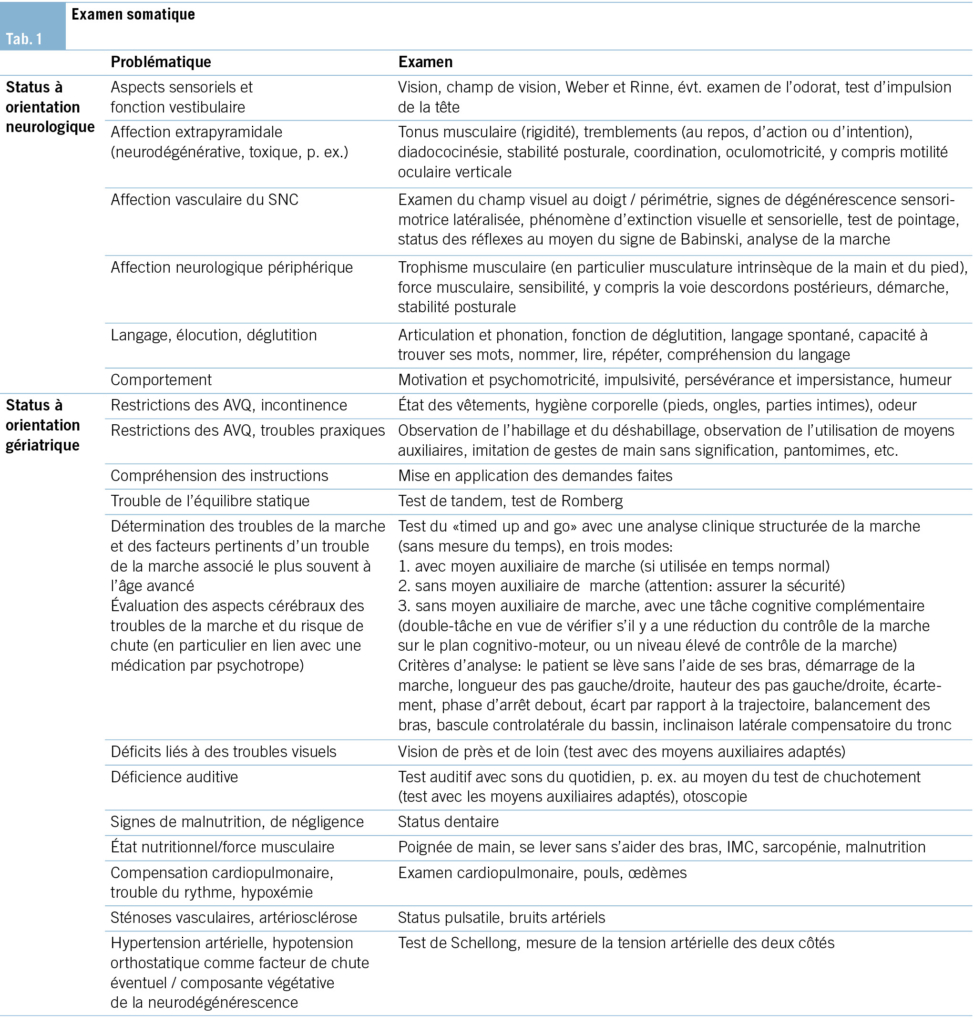
Examen somatique

L’ examen somatique est dirigé selon les comorbidités et le diagnostic suspecté (élément principal de la problématique) et doit être adapté en conséquence (Tab. 1).

Status psychopathologique et symptômes comportementaux et psychologiques de la démence (SCPD)

Outre les troubles cognitifs et les limitations dans les activités de la vie quotidienne, les patients atteints de démence présentent souvent, au cours de l’ évolution de la maladie, différents symptômes psychiatriques et comportementaux qui les handicapent de façon importante et compliquent leur traitement et leur prise en charge (12). Il peut s’ agir de troubles psychiques indépendants de la démence, ou reliés à celle-ci. Pour désigner les symptômes neuropsychiatriques se manifestant dans le cadre ou à la suite de la démence, le terme de «symptômes comportementaux et psychologiques de la démence» (SCPD) a été proposé. Pour l’ évaluation et le traitement, il est essentiel de prendre en compte les facteurs aussi bien biologiques et psychologiques qu’ environnementaux. À noter que les symptômes neuropsychiatriques peuvent apparaître avant le stade de la démence, au stade MCI (trouble cognitif léger) et peuvent même précéder les troubles cognitifs (13, 14). Les symptômes neuropsychiatriques se manifestant de façon précoce ont pour corollaire une aggravation plus rapide de la cognition et des fonctionnalités au quotidien (15).

Examen de base

• Évaluation clinique de la présence de symptômes psychiques
– Évaluation subjective par le patient
– Hétéroévaluation par les proches ou par des personnes connaissant bien le patient
– Examen psychopathologique de base
– Recherche d’ une éventuelle comorbidité psychiatrique, en particulier trouble dépressif, ou d’ un état confusionnel aigu
• Pondération des informations dans le processus de diagnostic
• Prise en compte des comorbidités psychiatriques primaires et des symptômes neuropsychiatriques/SCPD dans les propositions thérapeutiques relatives aux troubles cognitifs
• Transmission du cas vers une consultation spécialisée, en particulier lorsque les symptômes psychiatriques sont modérés à sévères, s’ il y a suspicion ou confirmation de comorbidités psychiatriques, s’ il y a des implications complexes pour la famille ou si la question d’ éventuelles mesures restreignant la liberté de mouvement se pose.

Optionnel

• L’ utilisation d’ une grille de description psychopathologique est recommandée (AMDP).
• L’ utilisation d’ échelles psychopathologiques spécifiques, p. ex. NPI-Q, est également recommandée. En cas de suspicion de dépression, l’ échelle de dépression gériatrique (GDS) (16) ou le Beck Depression Inventory (BDI-II) (17) sont également recommandés, si le patient se trouve encore au stade précoce de la démence.
• Traitement des comorbidités psychiques à la clinique de la mémoire: selon compétences et disponibilité

Examen neuropsychologique

Le principal bénéfice des tests cognitifs dans le cadre d’un examen neuropsychologique est le dépistage précoce et la quantification des dimensions cognitives altérées. Un profil des forces et faiblesses cognitives constitue en outre une base importante pour une psychoéducation, un conseil et un traitement adaptés aux besoins des patients et de leurs proches. À l’ instar de la présentation différenciée des troubles neurocognitifs du DSM-5 (18), l’ examen neuropsychologique complet vise à fournir des indications tant qualitatives que quantitatives sur les six dimensions suivantes:
• Attention: attention soutenue, attention divisée, attention sélective, vitesse de traitement
• Fonctions exécutives: planification, prise de décision, mémoire de travail, analyse des feedbacks/correction des erreurs, comportement différant des habitudes/inhibition comportementale, flexibilité mentale
• Apprentissage et mémoire: mémoire immédiate, mémoire à court terme (y c. rappel libre ou indicé et reconnaissance), mémoire à long terme (sémantique, autobiographique), apprentissage implicite
• Langage: production langagière (y c. dénomination et recherche de mots, fluence verbale, grammaire et syntaxe), compréhension
• Capacités perceptivo-motrices: capacité visuo-spatiales, visuo-constructives, perceptivo-motrices, praxies, gnosies
• Cognition sociale: reconnaissance des émotions, capacité d’ empathie (théorie de l’ esprit), changements de comportement
L’ évaluation des performances reposera sur:
• la capacité mesurée lors des tests (au moins deux méthodes de tests par domaine, p. ex. mémoire verbale et visuelle), ainsi que
• l’ observation/les données anamnestiques.
À l’ exception de la dimension «cognition sociale» (pour laquelle des instruments sont en cours de développement), il existe pour tous les aspects cognitifs des instruments de test qui ne nécessitent pas un investissement excessif en temps. Il s’ agit d’ utiliser des instruments qui définissent des normes prenant en compte l’ âge, le genre et la formation des personnes examinées.
Un tel examen standard sera complété par des instruments neuropsychologiques supplémentaires appropriés si le niveau cognitif initial est élevé, si des questions se posent à propos de l’ aptitude au travail, ainsi que pour les formes de démence rares ayant des modèles de référence cognitifs spécifiques.

Procédure pratique

Le standard minimum requis consiste à réaliser un bref test cognitif dont les résultats permettront de choisir la bonne option pour l’ examen neuropsychologique approfondi, compte tenu des informations tirées de l’ anamnèse, de l’ hétéro-anamnèse et en particulier des AVQ de base et des AIVQ. Le MoCA (7) ou le MMSE (5) combiné au test de l’ horloge (19) sont recommandés et font ainsi partie intégrante de la méthode standardisée. Un test cognitif différencié peut s’ avérer superflu pour les patients atteints de démence avancée.
Dans le cadre de l’ examen neuropsychologique, on appliquera dans un premier temps la méthode rapide, car elle permet de choisir la méthode et le niveau adéquats pour l’ examen neuropsychologique complet, en tenant compte des informations issues de l’ anamnèse et de l’ hétéro-anamnèse. Par la suite, une évaluation cognitive personnalisée et complète sera réalisée sur la base d’ hypothèses. Le résultat du test rapide, associé aux informations concernant l’ âge, le niveau de formation, les compétences linguistiques et le statut professionnel, permet de choisir une batterie appropriée de tests cognitifs complets, à l’ aide d’ un algorithme (Suppl. Fig.) (20). Pour les patients dont les capacités cognitives ne sont pas (encore) gravement altérées, l’ examen neuropsychologique approfondi constitue la norme dans les Memory clinic. En Suisse alémanique, le «Consortium to Establish a Registry for Alzheimer’ s Disease – Neuropsychological Assessment Battery (CERAD-NAB)», enrichi de tests mesurant la vitesse de traitement et les fonctions exécutives (CERAD-Plus), est une batterie de tests dûment standardisés et validés qui connaît un grand succès. Cette batterie est en revanche substituée en Suisse romande par une combinaison de tests cognitifs équivalents, dont la sélection est guidée par des hypothèses et constitue la base d’ un examen neuropsychologique personnalisé. Les éléments du test CERAD-Plus, y compris la possibilité de les évaluer, les tests utilisés en Suisse romande, ainsi qu’ une liste plus exhaustive de tests sont gratuitement à disposition sur le site www.swissmemoryclinics.ch/fr/.

Examen de groupes de patients particuliers

L’ examen est particulièrement complexe dans les cas suivants: (a) contexte d’ immigration avec compétences linguistiques limitées dans la langue de l’ examen (le cas échéant, il est judicieux de faire appel à un interprète interculturel, ce qui permet à long terme d’ épargner des ressources), (b) déficits sensoriels pour lesquels il n’ existe généralement pas de normes de référence, (c) troubles du développement intellectuel, (d) haut potentiel, (e) personnes très âgées (> 90 ans). Dans de telles conditions, l’ examen cognitif et son interprétation doivent s’ adapter à la situation individuelle et requièrent une vaste expertise neuropsychologique. En outre, il faut dans tous les cas tenir compte des troubles somatiques et psychiatriques ou des médicaments entravant les capacités cognitives, qui peuvent créer un biais dans l’ évaluation.

Évaluation et interprétation

La classification recommandée est celle que propose l’ Association suisse des neuropsychologues (ASNP) https://neuro.psychologie.ch/fr dans ses lignes directrices pour la classification et l’ interprétation des résultats aux tests neuropsychologiques, qui définit sept catégories. Le rapport sur l’ examen neuropsychologique devra indiquer en toute transparence le type de données sur lesquelles reposent les conclusions. En outre, il convient de prendre brièvement position sur le tableau clinique. Il est très utile de représenter dans un graphique les résultats des tests cognitifs sur la base des six dimensions cognitives selon le DSM-5. Il est encore judicieux et souhaitable – sur la base des résultats des tests cognitifs et de réflexions d’ ordre fonctionnel et neuro-anatomique – d’ envisager les hypothèses relatives à l’ étiologie d’ un trouble existant. Les réflexions sur l’ étiologie concrète et le diagnostic différentiel n’ ont évidemment de sens qu’ à condition d’ y intégrer toutes les données recensées à la clinique de la mémoire.

Diagnostic de laboratoire

Diagnostic sanguin

Examen de base
• Formule sanguine, protéine C-réactive
• Glucose
• Sodium, potassium, calcium corrigé
• Créatinine, eGFR
• TGO (transaminase glutamique oxalacétique), TGP (transaminase glutamique pyruvique), γ-GT (gamma-glutamyl transpeptidase)
• TSH (thyréostimuline)
• Vitamine B12, acide folique
• Cholestérol, cholestérol HDL, triglycérides (statut lipidique chez les moins de 80 ans)

Examens complémentaires en cas de suspicion spécifique ou de résultats pathologiques des examens de base
• Vitamine D
• Sérologies syphilis et borréliose, test VIH
• CDT (carbohydrate deficient transferrin)
• T3L, T4L, hormone parathyroïdienne, cortisol
• Numération sanguine différentielle, VS, INR
• CK (créatine kinase), urée, acide urique, bilirubine, phosphate, chlorure, magnésium, zinc
• Profil glycémique journalier, HbA1c
• Vitamine B1, vitamine B6, niacine, homocystéine, holotranscobolamine et/ou acide méthylmalonique
• Ferritine, transferrine
• Cuivre, céruloplasmine, status urinaire avec clairance du cuivre dans les urines de 24h
• Dépistage de substances toxiques (plomb, mercure)
• Dépistage de drogues (p. ex. benzodiazépines)
• Monitorage de drogues
• Anticorps des encéphalites auto-immunes ou paranéoplasiques
• Paramètres de vascularites
• Génotypage de l’ ApoE (p. ex. dans le cadre de la recherche, en cas de traitement anti-amyloïde prévu avec anticorps monoclonaux)

Diagnostic du liquide céphalo- rachidien (LCR)

Diagnostic standard pour les indications suivantes (21):
Exclusion des formes autres que les démences primaires dégénératives, en particulier ici les maladies inflammatoires chroniques du SNC.
• En présence de démences à progression rapide, atypiques ou précoces (première manifestation avant l’ âge de 65 ans).
• Ponction diagnostique ou de décharge en cas de suspicion d’ hydrocéphalie à pression normale.
• Diagnostic de soutien pour confirmer une neurodégénérescence, une pathologie tau et/ou une pathologie amyloïde ; s’ il y a une indication clinique spécifique et une investigation en cas de suspicion de la maladie d’ Alzheimer à un stade précoce (y compris le stade du trouble cognitif léger, MCI).
En cas de suspicion d’ une affection inflammatoire chronique du SNC, il convient, en plus du diagnostic de base du LCR, de déterminer des bandes oligoclonales et éventuellement des sérologies syphilis et borréliose, ainsi que d’ autres déterminations sérologiques. La recherche de l’ amyloïde bêta (Aβ42 ; Aβ42/Aβ40), de la protéine phospho-tau et de la protéine tau, en tant que biomarqueurs pour les démences, est appropriée comme diagnostic précoce et diagnostic différentiel dans l’ investigation des troubles cognitifs. Elle est généralement utilisée comme une partie du diagnostic de base dans le LCR. S’ il y a, cliniquement, une suspicion de la maladie de Creutzfeldt-Jakob, il est possible de déterminer en plus la protéine 14-3-3 et de faire une RT-QuIC (Real-Time Quaking-Induced Conversion) dans le LCR. Il est recommandé de procéder à une information individualisée des patients relative à l’ examen prévu des biomarqueurs et aux résultats possibles (21).

Imagerie diagnostique

Neuroradiologie

Les examens d’imagerie cérébrale réalisés dans le cadre du diagnostic des démences remplissent deux fonctions essentielles:
1. exclure les causes secondaires d’ une démence, comme la présence d’ une masse, les troubles de circulation du LCR, ainsi que les modifications d’ origine vasculaire, métabolique ou inflammatoire ;
2. contribuer à la différenciation étiologique et à la classification des formes primaires de démence.
L’ imagerie cérébrale peut contribuer au diagnostic et au diagnostic différentiel entre la maladie d’ Alzheimer et d’ autres démences, p. ex. fronto-temporales, bien qu’ à l’ heure actuelle, l’ imagerie structurelle ne suffit pas à elle seule à trancher entre ces étiologies (22). Cette imagerie est d’ une grande utilité pour identifier et évaluer les lésions intracérébrales ainsi que pour en estimer le nombre et la localisation. Ainsi, l’ imagerie doit être reconnue pour son apport à l’ évaluation globale et à la classification différentielle des tableaux cliniques des démences, en combinaison avec l’ anamnèse et les résultats cliniques, neuropsychologiques et d’ autres ordres.

Examen de base
L’ imagerie structurelle avec tomographie par résonance magnétique crânienne (IRM cérébrale) – ou à défaut, en cas de contre-indication à l’ IRM, par tomodensitométrie crânienne (CT) – fait partie du diagnostic de base.

Imagerie par résonance magnétique cérébrale
(IRM cérébrale)
Le choix se portera en général sur l’ IRM, notamment en raison de l’ absence d’ exposition au rayonnement et parce que la résolution des images anatomiques est bien meilleure qu’ avec un CT. Si cela est possible, on privilégiera une IRM 3 Tesla plutôt que 1,5 Tesla. Les recommandations détaillées sur le protocole d’ examen figurent sur le site de la SMC (https://www.swissmemoryclinics.ch/fr).

Tomodensitométrie cérébrale (scanner cérébral, CT)
À défaut d’IRM et en cas de contre-indication (p. ex. stimulateur cardiaque, implants numériques, claustrophobie majeure), on procédera à un scanner cérébral (CT). Le CT sans produit de contraste est en règle générale suffisant pour confirmer ou exclure la présence d’ une masse, d’ un hématome sous-dural ou d’ une hydrocéphalie et, avec certaines réserves, pour identifier une démence vasculaire.

Sonographie des vaisseaux irriguant le cerveau
En cas de démence vasculaire ou de formes mixtes de démence vasculaire et dégénérative, il peut être pertinent d’ évaluer les sténoses des vaisseaux cérébraux par échographie Doppler ou duplex.

Médecine nucléaire

Tomographie par émission de positons (TEP) au fluorodésoxyglucose (18F) (TEP–FDG)
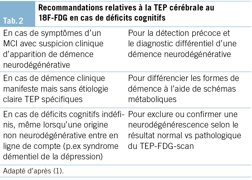
La TEP-FDG permet de représenter en images le métabolisme régional du glucose et de le comparer avec des ­valeurs normales. Il s’ agit de la technique d’ imagerie de médecine nucléaire la mieux validée pour le diagnostic des démences. Les recommandations relatives à la TEP-FDG figurent dans le Tab. 2.

La TEP-FDG présente une sensibilité élevée pour la mise en évidence de modifications fonctionnelles et neurodégénératives du traitement cortical des informations. Elle convient parfaitement au diagnostic précoce, même au stade de troubles cognitifs légers (MCI). La corrélation topique des aires présentant un dysfonctionnement permet par ailleurs, dans de nombreux cas, de faire la distinction entre la neurodégénérescence précoce fonctionnelle d’ états similaires à la démence (p. ex. en cas de maladie psychiatrique) et des troubles cognitifs d’ origine différente (p. ex. encéphalite limbique).
La TEP-FDG permet aussi de différencier des modèles métaboliques typiques de plusieurs autres démences neurodégénératives moins fréquentes, comme la démence à corps de Lewy et diverses formes de dégénérescences lobaires fronto-temporales– dont notamment les variantes de l’ aphasie primaire progressive (forme sémantique, non fluente et logopénique) et l’ atrophie corticale postérieure (23).
Pour résumer, la TEP-FDG est recommandée comme l’ imagerie moléculaire de premier choix pour le diagnostic des démences neurodégénératives (24). Pour une prise en charge des coûts selon l’ ordonnance sur les prestations de l’ assurance des soins (OPAS), des limitations sont à respecter.

TEP amyloïde
La TEP amyloïde permet de mettre en évidence avec certitude la présence ou l’ absence d’ une pathologie bêta-amyloïde dans le cerveau. Différents traceurs qui se lient aux plaques amyloïdes peuvent être utilisés. Depuis le 1er avril 2020, une obligation de prise en charge par les caisses maladie a été adoptée pour la TEP amyloïde comme examen complémentaire (limitations à respecter) notamment dans les cas peu clairs, après diagnostic du LCR non concluant ou lorsqu’ une ponction lombaire n’ est pas possible ou est contre-indiquée (24).
La mise à disposition prochaine de nouveaux médicaments actifs contre les pathologies amyloïdes élargira le champ d’ intervention clinique.

Imagerie du système dopaminergique: SPECT du transporteur de la dopamine avec l’ ioflupane (123I) et TEP à la 18F-DOPA
Le SPECT des transporteurs de la dopamine à l’ ioflupane (123I) (DaTSCAN®) et la TEP à la 18F-DOPA sont des examens de médecine nucléaire destinés à déterminer la disponibilité cérébrale de la dopamine (25). La mise en évidence d’ un déficit dopaminergique dans le striatum par ces examens permet de distinguer une maladie à corps de Lewy (avec résultat pathologique dans le striatum) et les démences sans corps de Lewy (en particulier Alzheimer) avec résultat normal dans le striatum (26). Swissmedic a autorisé deux produits radiopharmaceutiques pour le SPECT (DaTSCAN® et Striascan®) ainsi que la TEP à la 18F-DOPA pour cette indication.

Analyse génétique

Les personnes atteintes d’ une démence et leurs proches s’ inquiètent souvent de savoir si la maladie est héréditaire. Or il n’ est pas toujours nécessaire, au plan médical, de procéder à des analyses génétiques pour répondre à cette préoccupation (27).

Examen de base
Les professionnels d’ une clinique de la mémoire devront pouvoir:
• identifier les cas de démence familiale,
• peser l’ information dans le cadre de l’établissement du diagnostic,
• poser l’ indication d’ une consultation génétique spécialisée,
• collaborer avec les spécialistes en génétique humaine et documenter cette collaboration,
• discuter avec le patient/les proches de l’ éventuelle annonce vers une consultation en médecine génétique humaine,
• adresser le patient/la famille vers un institut de médecine génétique humaine (habituellement relié à une clinique universitaire). Cette procédure aura notamment lieu dans les cas suivants:
– un apparenté de 1er degré potentiellement atteint et âgé de moins de 50 ans
– deux apparentés de 1er degré potentiellement atteints et âgés de moins de 60 ans
• renseigner et conseiller à propos de la génétique en tant que facteur de risque des pathologies démentielles les plus fréquentes.

Autres examens

S’ appuyant sur des éléments anamnestiques ou des résultats de l’ examen clinique, les examens complémentaires suivants peuvent être appropriés et utiles.

Électroencéphalogramme (EEG)

Une EEG peut fournir des informations utiles dans les cas suivants:
• fluctuations importantes dans la vigilance et l’ orientation, en vue d’ exclure une origine épileptique,
• suspicion de maladie inflammatoire/infectieuse, inflammatoire/auto-immune ou métabolique du SNC (p. ex. encéphalite limbique, encéphalopathie de Hashimoto) et suspicion de la maladie de Creutzfeldt-Jakob.

Diagnostic d’ un trouble du sommeil

Les démences sont souvent accompagnées d’ un besoin accru de sommeil, autrement dit d’ un sommeil nocturne prolongé et d’ une somnolence diurne excessive. Tout comme les maladies organiques du cerveau, les troubles respiratoires associés au sommeil peuvent perturber l’ architecture du sommeil et induire une somnolence diurne excessive, ainsi qu’ une baisse de l’ attention. Ils représentent un facteur de risque indépendant pour des événements cardio-vasculaires et cérébro-vasculaires et répondent souvent aux traitements.
Une somnolence diurne excessive peut être évaluée simplement, au moyen du questionnaire de somnolence d’ Epworth (28). Un examen au cours du sommeil au moyen d’ un appareil (actimétrie, pulsoxymétrie nocturne, polygraphie, polysomnographie) peut être indiqué en cas de:
• somnolence diurne excessive et signes de troubles respiratoires nocturnes, en vue d’ exclure un syndrome d’ apnée du sommeil.
• suspicion d’ un trouble du sommeil paradoxal;
• suspicion de crises d’ épilepsie liées au sommeil.

Test de l’ odorat

Un test de l’ odorat peut fournir un résultat corroborant le diagnostic préliminaire lors du dépistage précoce de la maladie d’ Alzheimer Dans la maladie d’ Alzheimer ainsi que dans la maladie de Parkinson une baisse de l’ odorat peut apparaitre très tôt, accompagnant ou précédent les premiers troubles cognitifs. Différents tests validés sont à disposition.

Analyse de la marche

Outre la cognition, les pathologies neurodégénératives peuvent altérer les fonctions motrices. Les personnes affectées par une démence présentent un risque de chute accru par rapport aux sujets du même âge en bonne santé. Une analyse clinique structurée permet de déterminer l’ assurance à la marche et les risques de chute. Elle aide à poser l’ indication de mesures thérapeutiques ou préventives adéquates (entraînement, moyen auxiliaire de marche, etc.). L’ analyse quantitative de la marche repose sur des tests cliniques validés de la mobilité (p. ex. test «timed up and go»), mais aussi sur des procédés informatisés (les données de mobilité sont saisies numériquement, p. ex. au moyen de capteurs de pression sur un tapis de marche).

Oculomotricité/champ visuel

Un examen clinique de l’ oculomotricité et du champ visuel s’ impose en cas de suspicion de certaines maladies neurodégénératives (p. ex. paralysie supranucléaire progressive) ou de pathologie vasculaire.

Entretien d’annonce du diagnostic

L’ entretien d’annonce du diagnostic fait suite à la pose interdisciplinaire du diagnostic et constitue le point de départ d’ un conseil, d’ un traitement et d’ un accompagnement individualisés. De façon générale, le patient a le droit d’ être informé de manière claire et appropriée sur son état de santé, à moins qu’ il ne renonce explicitement à cette information (29). Étant donné la gravité du diagnostic, celui-ci doit être révélé avec le plus grand soin. L’ emploi de mots simples et clairs, le cas échéant adaptés aux limitations cognitives du patient, facilite la compréhension du diagnostic et sa portée et permet par la suite une communication ouverte au sein de la famille ou du cercle de référence. Il convient de donner au patient et à ses proches suffisamment de possibilités de poser des questions (30). En cas de besoin, un nouvel entretien sera proposé pour compléter le premier. Donner des informations sur les offres de soutien et d’ accompagnement pour les patients et les proches, ou les proposer déjà, peut représenter un soulagement important et contribuer à une meilleure maîtrise de la situation. Il est recommandé de fournir du matériel d’ information aux patients et aux proches, mais aussi de faire connaître les conseils et l’ accompagnement proposés par l’ organisation Alzheimer Suisse sur tout le territoire national.

Aspects particuliers

Lorsqu’ il y a une limitation des fonctions cognitives, les questions d’ une aptitude à la conduite ou d’ une capacité de discernement peuvent se poser(31). Dans le cadre de ses examens, la clinique de la mémoire ne propose pas à proprement parler d’ examen d’ aptitude à la conduite. En cas de limitation des performances cognitives ou comportementales, il peut cependant s’ avérer important de se préoccuper de leur pertinence par rapport à l’ aptitude à la conduite et, le cas échéant, d’ engager les démarches qui s’ imposent.
Si la capacité de discernement (32) doit être déterminée, cela se fera lors d’ une évaluation à part. Dans le cadre de l’ entretien d’ annonce du diagnostic, il est toutefois recommandé d’ évoquer la problématique avec les personnes concernées au stade précoce de la maladie déjà, et de leur suggérer de se faire conseiller sur les plans juridique, financier et médical afin de pouvoir prendre au besoin, de leur propre chef, les mesures nécessaires (p. ex. mandat pour cause d’ inaptitude, directives anticipées).

Suite des démarches et suivi

En lien avec l’ entretien d’annonce du diagnostic, et à côté des recommandations liées aux mesures médicamenteuses et non médicamenteuses (10), il est conseillé de donner des informations sur les services sociaux ou les services de conseil relatifs à la vie de tous les jours, sur les groupes d’ entraide ainsi que sur les possibilités concrètes d’ accompagnement et de décharge disponibles dans la région. Le site www.alzguide.ch tient un registre national de telles offres. Par ailleurs, selon le pronostic et les besoins individuels, il peut être bon de planifier un suivi de l’ évolution ou un accompagnement de longue durée. Un tel suivi à la clinique de la mémoire permet de refaire le point sur le diagnostic, de conseiller et le cas échéant, d’ adapter le traitement et l’ accompagnement. Ces démarches devraient être engagées d’ entente avec les personnes concernées, leurs proches et les services médicaux, thérapeutiques et soignants, ainsi qu’ avec les entités spécialisées.

Perspective

Bien que la Suisse possède un réseau relativement dense de cliniques de la mémoire spécialisées (33) et que la pose précise et précoce du diagnostic soit recommandée (21, 34), la proportion de personnes atteintes de démence et ne bénéficiant pas d’ un diagnostic ou d’ un diagnostic suffisamment précis reste élevée. De plus, davantage d’ efforts sont nécessaires dans la recherche sur les aspects spécifiques aux hommes et aux femmes ainsi que pour l’ implémentation de méthodes et de processus diagnostiques qui tiennent compte de ces aspects (35). De même, il s’ agirait d’ améliorer les méthodes d’ évaluation des symptômes et déficits fonctionnels parmi des groupes spécifiques de patients, comme les patients affectés à un âge jeune ou moyen, les patients issus de différents horizons culturels, en particulier les migrants, ou d’autres groupes de patients.
Les progrès réalisés dans le diagnostic clinique, neuropsychologique et celui des biomarqueurs, ainsi que les méthodes numériques de saisie des symptômes devraient améliorer et élargir de façon considérable, dans les années à venir, la pratique clinique en lien avec l’ établissement de profils de risques individuels. Ces améliorations faciliteront tant le dépistage précoce que le monitorage des troubles cognitifs et démences. En outre, les processus neurocognitifs, les activités de la vie quotidienne et les symptômes neuropsychiatriques pourront être saisis et interprétés de façon plus précise dans l’ environnement quotidien. Ces développements offrent la perspective d’ un diagnostic plus précis et en même temps davantage lié au quotidien, ce qui facilitera un conseil et un accompagnement individualisés et permettra des interventions répondant mieux aux réels besoins. Les nouveaux traitements visant les pathologies cérébrales qui seront probablement bientôt disponibles, comme les anticorps anti-amyloïdes, requerront une identification précise, basée sur les biomarqueurs, de la pathologie visée ainsi qu’ une appréciation précise des risques liés au traitement.
Si ces innovations peuvent constituer un grand progrès, leur mise en œuvre s’ accompagne aussi de grands défis. En font partie les questions sur les aspects éthiques, l’ adaptation de la prise en charge, la disponibilité des ressources, ainsi que l’ acceptation et l’ application appropriée des nouvelles possibilités.

Prof. Dr. med. Julius Popp
University of Zürich
Department of Adult Psychiatry and Psychotherapy
Lenggstrasse 31, CH-8032 Zürich
julius.popp@uzh.ch

Julius Popp 1, 2*, Tatjana Meyer-Heim 1, 3, Markus Bürge 1, 3, Michael M. Ehrensperger 1, 4,
Ansgar Felbecker 1, 5, Hans Pihan 1, 5, 6, Nadège Barro-Belaygues 1, 3, 6, Christian Chicherio 1,
Bogdan Draganski 1, 5, Gaby Bieri 1, 3, Irene Bopp-Kistler 3, 7, Andrea Brioschi 8, Matthias Brühlmeier 9, Karin Brügger 1, 4, Gianclaudio Casutt 4, 10, 11, Valentina Garibotto 9, Petra Gasser 4,
Dan Georgescu 1, 2, Anton Gietl 12, Andrea Grubauer 4, Freimut Jüngling 9, Sonja Kagerer 12,
Eberhard Kirsch 13, Vera Kolly 4, Gabriela Latour Erlinger 1, 4, 12, Andreas Monsch 4, Laura Rinke 1, 4, Rafael Meyer 1, 2, 14

1 Swiss Memory Clinics (SMC)
2 Société suisse de psychiatrie et psychothérapie de la personne âgée (SPPA)
3 Société professionnelle suisse de gériatrie (SPSG)
4 Association suisse des neuropsychologues (ASNP)
5 Société suisse de neurologie (SSN)
6 Centre hospitalier Bienne
7 mediX Gruppenpraxis Zürich
8 Centre hospitalier universitaire vaudois (CHUV)
9 Société suisse de médecine nucléaire (SSMN)
10 Société suisse de psychologie de la circulation (SPC)
11 Swiss Insurance Medicine (SIM)
12 Psychiatrische Universitätsklinik Zürich (PUK)
13 Société suisse de neuroradiologie (SSNR)
14 Psychiatrische Dienste Aargau AG (PDAG)

Abréviations
AD Alzheimer’ s disease, maladie d’ Alzheimer
AIVQ activités instrumentales de la vie quotidienne
AVQ activités de la vie quotidienne
CT computed tomography, scanner
IRM Imagerie par résonance magnétique
MCI Mild Cognitive Impairment
MMSE Mini-Mental-Status-Examination
MoCA Montreal-Cognitive-Assessment
SCPD symptômes comportementaux et psychologiques de la démence
SMC Swiss Memory Clinics
TEP tomographie par émission de positons

Historie
Manuscrit soumis: 24.02.2025
Manuscrit accepté: 03.03.2025

Prof. Dr. med. Julius Popp

Swiss Memory Clinics (SMC)
Schweizerische Gesellschaft für Alterspsychiatrie (SGAP)

Department of Adult Psychiatry and Psychotherapy
University of Zürich
Lenggstrasse 31
CH-8032 Zürich

Die Autorenschaft hat keine Interessenkonflikte im Zusammenhang mit diesem Artikel deklariert.

• Die aktualisierten Empfehlungen bilden den heutigen Stand der vorhandenen Diagnosemöglichkeiten in der Schweiz ab.
• Die Empfehlungen sollen der Qualitätsentwicklung sowie als Wegweiser für den klinischen Alltag dienen.
• Ein wesentliches Ziel ist, die Früh- und Differenzialdia­gnostik der Demenzerkrankungen zu verbessern.

1. Bürge M, Bieri G, Brühlmeier M, Colombo F, Demonet JF, Felbecker A, et al. [Recommendations of Swiss Memory Clinics for the Diagnosis of Dementia]. Praxis (Bern 1994). 2018;107(8):435-51.
2. Bundesamt für Gesundheit BAG, Swiss Memory Clinics. Therapieempfehlungen Demenz 2024 [Available from: https://www.bag.admin.ch/dam/bag/de/dokumente/nat-gesundheitsstrategien/nationale-demenzstrategie/hf-angebote/3_4_akutspital/therapieempfehlungen-demenz.pdf.download.pdf/broschuere-therapieempfehlungen-demenz.pdf.
3. Klöppel S, Meyer-Heim T, Ehrensperger M, Rüttimann A, Weibel I, Schnelli A, et al. [The Swiss Memory Clinics recommendations for the treatment of dementia]. Praxis (Bern 1994). 2024;113(8):187-94.
4. Ranson JM, Kuzma E, Hamilton W, Lang I, Llewellyn DJ. Case-finding in clinical practice: An appropriate strategy for dementia identification? Alzheimers Dement (N Y). 2018;4:288-96.
5. Moyer VA, Force USPST. Screening for cognitive impairment in older adults: U.S. Preventive Services Task Force recommendation statement. Ann Intern Med. 2014;160(11):791-7.
6. Folstein MF, Folstein SE, McHugh PR. “Mini-mental state”. A practical method for grading the cognitive state of patients for the clinician. J Psychiatr Res. 1975;12(3):189-98.
7. Thalmann B, Spiegel R, Monsch A, Stähelin H, Brubacher D, Ermini-Funfschilling D, et al. Dementia screening in general practice: optimised scoring for the clock drawing test. Brain Aging Int J. 2002;2:36-43.
8. Kalbe E, Kessler J, Calabrese P, Smith R, Passmore AP, Brand M, et al. DemTect: a new, sensitive cognitive screening test to support the diagnosis of mild cognitive impairment and early dementia. Int J Geriatr Psychiatry. 2004;19(2):136-43.
9. Nasreddine ZS, Phillips NA, Bédirian V, Charbonneau S, Whitehead V, Collin I, et al. The Montreal Cognitive Assessment, MoCA: a brief screening tool for mild cognitive impairment. J Am Geriatr Soc. 2005;53(4):695-9.
10. Lawton MP, Brody EM. Assessment of older people: self-maintaining and instrumental activities of daily living. Gerontologist. 1969;9(3):179-86.
11. Sheikh K, Smith DS, Meade TW, Goldenberg E, Brennan PJ, Kinsella G. Repeatability and validity of a modified activities of daily living (ADL) index in studies of chronic disability. Int Rehabil Med. 1979;1(2):51-8.
12. Jekel K, Damian M, Wattmo C, Hausner L, Bullock R, Connelly PJ, et al. Mild cognitive impairment and deficits in instrumental activities of daily living: a systematic review. Alzheimers Res Ther. 2015;7(1):17.
13. Lyketsos CG, Carrillo MC, Ryan JM, Khachaturian AS, Trzepacz P, Amatniek J, et al. Neuropsychiatric symptoms in Alzheimer’s disease. Alzheimers Dement. 2011;7(5):532-9.
14. Sheikh F, Ismail Z, Mortby ME, Barber P, Cieslak A, Fischer K, et al. Prevalence of mild behavioral impairment in mild cognitive impairment and subjective cognitive decline, and its association with caregiver burden. Int Psychogeriatr. 2018;30(2):233-44.
15. Ismail Z, Smith EE, Geda Y, Sultzer D, Brodaty H, Smith G, et al. Neuropsychiatric symptoms as early manifestations of emergent dementia: Provisional diagnostic criteria for mild behavioral impairment. Alzheimers Dement. 2016;12(2):195-202.
16. Peters ME, Schwartz S, Han D, Rabins PV, Steinberg M, Tschanz JT, et al. Neuropsychiatric symptoms as predictors of progression to severe Alzheimer’s dementia and death: the Cache County Dementia Progression Study. Am J Psychiatry. 2015;172(5):460-5.
17. Kaufer DI, Cummings JL, Ketchel P, Smith V, MacMillan A, Shelley T, et al. Validation of the NPI-Q, a brief clinical form of the Neuropsychiatric Inventory. J Neuropsychiatry Clin Neurosci. 2000;12(2):233-9.
18. Yesavage JA, Brink TL, Rose TL, Lum O, Huang V, Adey M, et al. Development and validation of a geriatric depression screening scale: a preliminary report. J Psychiatr Res. 1982;17(1):37-49.
19. BECK AT, WARD CH, MENDELSON M, MOCK J, ERBAUGH J. An inventory for measuring depression. Arch Gen Psychiatry. 1961;4:561-71.
20. Falkai P, Wittchen H-U. Diagnostisches und statistisches Manual psychischer Störungen DSM-5. 2 ed. (APA) APA, editor. Göttingen: Hogrefe; 2018.
21. Popp J, Georgescu D, Bürge M, Mundwiler-Pachlatko E, Bernasconi L, Felbecker A. [Biomarkers for the diagnosis of cognitive impairment – Recommendations from the Swiss Memory Clinics]. Praxis (Bern 1994). 2022;111(13):738-44.
22. Krueger CE, Dean DL, Rosen HJ, Halabi C, Weiner M, Miller BL, et al. Longitudinal rates of lobar atrophy in frontotemporal dementia, semantic dementia, and Alzheimer’s disease. Alzheimer Dis Assoc Disord. 2010;24(1):43-8.
23. Juengling FD, Allenbach G, Bruehlmeier M, Klaeser B, Wissmeyer MP, Garibotto V, et al. Appropriate use criteria for dementia amyloid imaging in Switzerland – mini-review and statement on behalf of the Swiss Society of Nuclear Medicine and the Swiss Memory Clinics. Nuklearmedizin. 2021;60(1):7-9.
24. Guedj E, Varrone A, Boellaard R, Albert NL, Barthel H, van Berckel B, et al. EANM procedure guidelines for brain PET imaging using [Eur J Nucl Med Mol Imaging. 2022;49(2):632-51.
25. Morbelli S, Esposito G, Arbizu J, Barthel H, Boellaard R, Bohnen NI, et al. EANM practice guideline/SNMMI procedure standard for dopaminergic imaging in Parkinsonian syndromes 1.0. Eur J Nucl Med Mol Imaging. 2020;47(8):1885-912.
26. Papathanasiou ND, Boutsiadis A, Dickson J, Bomanji JB. Diagnostic accuracy of ¹²³I-FP-CIT (DaTSCAN) in dementia with Lewy bodies: a meta-analysis of published studies. Parkinsonism Relat Disord. 2012;18(3):225-9.
27. Johns MW. A new method for measuring daytime sleepiness: the Epworth sleepiness scale. Sleep. 1991;14(6):540-5.
28. Bundesamt für Gesundheit BAG. Das Recht auf Aufklärung 2020 [Available from: https://www.bag.admin.ch/bag/de/home/medizin-und-forschung/patientenrechte/rechte-arzt-spital/1-recht-aufklaerung.html.
29. Bopp-Kistler I. [Disclosing the diagnosis and guidance]. Ther Umsch. 2015;72(4):225-31.
30. Meyer R, Monsch A, Allali G, Barro-Belaygues N, Becker S, Bürge M, et al. Alzheimer-Krankheit – Systembereitschaft im Kontext neuer Entwicklungen Praxis (Bern 1994). 2024;113(11&12):293–6.
31. Hemmeter U, Strnad J, Decrey-Wick H, Christina Affentranger C, Bättig E, Becker S, et al. Weiterentwicklung von Empfehlungen in den Bereichen Früherkennung, Diagnostik und Behandlung für die Grundversorgung 2021 [Available from: https://www.bag.admin.ch/dam/bag/de/dokumente/nat-gesundheitsstrategien/nationale-demenzstrategie/hf-qualitaet/6_1_grundversorgung/Empfehlungen_Grundversorgung%20.pdf.download.pdf/Empfehlungen%20in%20den%20Bereichen%20Früherkennung,%20Diagnostik%20und%20Behandlung%20für%20die%20Grundversorgung.pdf.
32. Aggarwal NT, Mielke MM. Sex Differences in Alzheimer’s Disease. Neurol Clin. 2023;41(2):343-58.

Antibiotic Prophylaxis in Gastrointestinal Endoscopy

Introduction

The need for antibiotic prophylaxis for endoscopic procedures, especially in the gastrointestinal tract, has long been a matter of debate. In recent years, the lack of randomized trials supporting a benefit of antibiotic prophylaxis, the very low incidence of infective endocarditis after endoscopic procedures and the potential adverse reactions of antibiotics have led to a more restricted use of antibiotic prophylaxis.

The aim of this expert opinion statement is to provide an overview of current evidence and to propose a pragmatic approach to the use of antibiotic prophylaxis for endoscopic procedures in areas where clear evidence is lacking.

All recommended antibiotic regimens are summarized in Tab. 1.

The GRADE system is used for rating the quality of evidence.

High-quality evidence: recommendation grade A
Evidence comes from one or more well-designed and well-executed randomized controlled trials that yield consistent and directly applicable results. Further research is very unlikely to change our confidence in the estimate of effect.

Moderate-quality evidence: recommendation grade B
Evidence comes from randomized controlled trials with important limitations and a very small number of participants, well-designed controlled trials without randomization, well-designed cohort or case-control studies, and multiple time series with or without intervention. Further research will probably have an important impact on our confidence in the estimate of effect and may change the estimate.

Low-quality evidence: recommendation grade C
Evidence comes from observational studies. Further research is very likely to have an important impact on our confidence in the estimate of effect and will probably change the estimate.

Very-low quality evidence: recommendation grade D
Evidence is conflicting, of poor quality, or lacking, and hence the balance of benefits and harms cannot be determined. Any estimate of effect is very uncertain as evidence is either unavailable or does not permit a conclusion.

Risk of bacteremia

Transient bacteremia is very common during many routine daily activities, such as tooth brushing (up to 68 %), using toothpicks (up to 40 %), or even chewing food (up to 50 %). These numbers are essential to consider when evaluating the incidence of transient bacteremia associated with gastrointestinal procedures.

Gastroscopy, flexible sigmoidoscopy and colonoscopy are all considered low-risk procedures for bacteremia and infection, regardless of whether biopsies are taken or polypectomies are performed. Mean rates of bacteremia were estimated to be 1 % in sigmoidoscopy and 4 % in gastroscopy and colonoscopy, whereas esophageal dilation carries the highest risk for bacteremia. (Tab. 2)

Endoscopic procedures with high-risk for ­bacteremia

ERCP in an obstructed bile duct
The risk for bacteremia increases from 6 % in the absence of biliary obstruction to 18 % with obstruction (2). Therefore, in cases of biliary obstruction the incidence of post-ERCP cholangitis is increased.

Esophageal dilation
Bacteremia during or after esophageal dilation is found in up to 22 % of patients and may be higher in the case of multiple passes and/or in patients with malignant stenosis (3, 4).

Sclerotherapy of varices
The range of reported bacteremia rates is wide, spanning between 4 % and 56 % in different studies, with an average rate around 20 % in most studies (5).

In contrast, rubber band ligation of esophageal varices is not considered a high-risk procedure for bacteremia, with an estimated risk 9 % (6).

Risk of infective endocarditis after ­endoscopic procedures

Although bacteremia, as outlined above, is a common event during endoscopic procedures, subsequent infective endocarditis (IE) is extremely rare. Despite the constantly rising number of endoscopic procedures performed worldwide, there has been no evidence of an increasing incidence of IE after such procedures.

Furthermore, only limited data exist regarding the impact of antibiotic prophylaxis for dental or surgical procedures on the prevention of IE. Failures of endocarditis prophylaxis, despite the correct administration of antibiotic prophylaxis, are well recognized (7). For these reasons, antibiotic prophylaxis is generally not recommended to reduce the incidence of infective endocarditis in GI endoscopic procedures.

Patients at high risk of infective endocarditis

In the recently published guideline of the European Society of Cardiology, there is a novel recommendation for patients at high risk of infective endocarditis (Tab. 3), stating that “antibiotic prophylaxis may be considered for high-risk patients undergoing an invasive diagnostic or therapeutic procedure in the gastrointestinal and genitourinary tract, skin or musculoskeletal system” (8).

There is no evidence supporting the use of antimicrobial prophylaxis for cardiac transplant recipients who develop cardiac valvulopathy. The indication should be discussed on a case-by-case basis. Patients should contact their transplant specialist to evaluate the indication prior to an elective intervention.
To prevent infective endocarditis, antibiotic prophylaxis may be considered in high-risk patients undergoing an invasive diagnostic or therapeutic procedure in the GI tract (Tab. 2) (low quality evidence).

In patients at high risk of IE, the following antibiotic regimen is recommended (9). (Tab. 4)

Endoscopic procedures due to infective ­disorders in high-risk patients of infective endocarditis

In the case of an established infection likely caused by enterococci, an empiric antibiotic regimen with anti-enterococcal activity should be used. These patients, if already receiving antibiotic therapy, do not need additional antibiotic prophylaxis when undergoing an endoscopic procedure (Tab. 3).

Specific endoscopic procedures: When to use antibiotic prophylaxis

Routine upper endoscopy and colonoscopy

Antibiotic prophylaxis is not required in routine gastrointestinal endoscopy including biopsies and polypectomy, even if high-risk procedures are performed during routine endoscopy (Tab. 2). Exceptions may include patients with severe neutropenia (< 500 cells/mm3) and advanced hematologic malignancies (35).

Colorectal endoscopic mucosal resection (EMR) and endoscopic submucosal dissection (ESD)

EMR and ESD are frequently performed to treat benign and early malignant colorectal lesions. However, infections following EMR and ESD are extremely rare.

Routine use of antibiotic prophylaxis is therefore not recommended.

Post-EMR/ESD Coagulation Syndrome (PECS)
In the context of large colorectal endoscopic resections, a novel complication known as “post-EMR/ESD coagulation syndrome” (PECS) has been recognized. The syndrome is characterized by pain, local peritonitis, fever and elevation of inflammatory markers. It occurs in up to 40 % of patients undergoing a large endoscopic resection.

Large lesions (>30 mm) and localization outside the rectosigmoid colon are independent risk factors for the development of PECS. Limited data are available on the effect of antibiotic prophylaxis in such patients. In one randomized controlled trial of 409 patients (randomized to either cefuroxime 1.5 g iv half an hour before and 6 hours after the intervention or placebo), the rate of adverse events in the antibiotic group was significantly lower than in the control group: abdominal pain (2.8 % vs 14.9 %, p < 0.01), diarrhea (2.0 % vs 9.3 %, p < 0.05), and fever (0.9 % vs 8.4 %, p < 0.05), respectively. The levels of inflammatory markers were also significantly lower in the antibiotic group compared with those in the control group (11). Therefore, antibiotic prophylaxis can be applied to reduce the risk of PECS in patients with large endoscopic resections above the rectosigmoid colon.
To prevent PECS, antibiotic prophylaxis can be considered in patients with large colonic endoscopic resections above the sigmoid colon (low-quality evidence).

Endoscopic retrograde cholangio-pancreatography (ERCP)

Cholangitis is the most common infectious adverse event in ERCP. Others are cholecystitis, duodenoscope-related transmission of infections, and endocarditis (12).

The role of antibiotic prophylaxis in reducing the risk of post-ERCP cholangitis has been evaluated in several studies. The most recent a meta-analysis from 2010 (9 RCTs with 1573 patients) (13) found a lower risk of post-ERCP cholangitis. In the subgroup of patients who were drained after the first ERCP, there was no benefit from antibiotic prophylaxis.

Subsequent studies did not find a benefit of antibiotic prophylaxis except in patients with biliary obstruction. A very recent randomized trial of 378 patients found a significantly reduced rate of infectious complications, especially cholangitis (2.8 % vs 9.8 % p = 0.007) in patients with biliary obstruction who received antibiotic prophylaxis (14).
Antibiotic prophylaxis is not recommended if cholangitis is absent, if biliary drainage is likely to be successful, and in patients undergoing ERCP for reasons other than biliary obstruction (high-quality evidence).

Malignant hilar obstruction and PSC
Especially in patients with malignant hilar obstruction and primary sclerosing cholangitis, the risk of unsuccessful drainage is higher, which raises the risk of complicating cholangitis (15).

Peroral cholangioscopy
Several studies have found that peroral cholangioscopy is associated with a high risk of bacteremia and cholangitis, regardless of the indication for the intervention. For this reason, all patients undergoing peroral cholangioscopy should receive antibiotic prophylaxis (16).
To prevent post-ERCP cholangitis and/or sepsis, antibiotic prophylaxis is recommended for patients with biliary obstruction, PSC, and in patients undergoing cholangioscopy (moderate-quality evidence).

In the case of failed biliary drainage, antibiotic therapy may be continued for 3–5 days (17).

Endosonographic-guided tissue acquisition

EUS-guided puncture/biopsy is an important, minimally invasive technique for obtaining tissue diagnoses from a variety of pancreatic, intraabdominal, retroperitoneal, or mediastinal lesions in close proximity to the gastrointestinal tract. The major complications associated with EUS-FNA include hemorrhage, perforation, infection, and organ-specific complications, such as acute pancreatitis following puncture of pancreatic lesions. A previous systematic review of complications and deaths associated with EUS-FNA (51 reports, 10 941 patients) revealed an overall complication rate of 0.98 %. The risk of infection was very low, at 0.05 % (18). The risk of infection depends on the type of lesion being sampled. Data suggest that solid lesions carry a very low risk for infection (0.01 %–2 %). The risk of infection following EUS-FNA of cystic lesions is less clear, with reported rates of infection ranging from < 1 % to 14 % (19).

Pancreatic lesions

Solid lesions of the pancreas
EUS-FNA and EUS-FNB carry a low risk of bacteremia and sepsis, especially in solid lesions. In two large series involving 627 patients undergoing EUS-FNA for a variety of solid lesions of the pancreas, sepsis developed in only 3 patients.
Antibiotic prophylaxis is not recommended before EUS-FNA/B of solid lesions of the pancreas (moderate-quality evidence).

Cystic lesions of the pancreas
It has been a long-standing practice to administer antibiotic prophylaxis in patients undergoing EUS-FNA of cystic pancreatic lesions, since older data suggest that this intervention is associated with higher rates of infection and antibiotic prophylaxis appeared to be efficient in such patients.

However, newer data do not support this practice and the role of antibiotic prophylaxis has been questioned. A meta-analysis from 2020 (six studies, including one randomized controlled trial and five retrospective studies, with 1706 patients) evaluated the efficacy of antibiotic prophylaxis prior to EUS-FNA of cystic pancreatic lesions. Overall, 8 infectious events were observed in the antibiotic group (0.77 %), and 12 events in the control group (1.7 %), (odds ratio (OR) 0.65, 95 % confidence interval (95 % CI) 0.24–1.78; p = 0.40). No difference was observed between the two study groups in terms of either severe infection (OR 0.88, 95 % CI 0.13–5.82; p = 0.89) or overall adverse event rate (OR 1.09, 95 % CI 0.73–1.65; p = 0.67). These findings suggest prophylactic antibiotics do not substantially reduce the risk of infections after EUS-FNA of cystic lesions of the pancreas (20).

One randomized trial from Spain compared the effect of antibiotic prophylaxis versus placebo in patients undergoing EUS-FNA of cystic lesions of the pancreas. More than 200 patients were randomly assigned to prophylaxis with ciprofloxacin (n = 112) or saline solution (n = 114, placebo). The only case of FNA-related infection (0.44 %) occurred in a patient in the placebo group (0.87 %). Prevention of infection was not inferior in the control group, and there were no differences between groups regarding the occurrence of post-interventional fever or other adverse events (21).
We suggest that antibiotic prophylaxis can be retained in patients undergoing EUS-FNA of cystic lesions of the pancreas and may be reserved for special situations (moderate-quality evidence)

Rectal and perirectal lesions

EUS-guided transrectal tissue acquisition is a safe technique to obtain tissue diagnosis of solid perirectal lesions. The infection rate after such interventions is very low.

In a prospective study, 100 patients underwent a total of 471 fine needle aspirations of rectal or perirectal lesions. Blood cultures were taken in all patients before and after the intervention. Of these, cultures were positive in 6 patients and 4 patients had contamination. Two patients developed bacteremia with either Bacteroides fragilis or Gemella morbillorum. No signs or symptoms of infection developed in any patient.
Therefore, EUS-FNA of solid lesions in the lower GI tract can be considered a low-risk procedure for infection and does not warrant antibiotic prophylaxis (22).
Antibiotic prophylaxis is not recommended in patients undergoing EUS-FNA of solid perirectal lesions (moderate-quality evidence).

Mediastinal lesions

EUS-guided transesophageal puncture is a safe technique that allows tissue acquisition for diagnosis of undetermined mediastinal lesions. Special caution is required with cystic lesions, as post-interventional infection of such lesions may be life threatening.

Solid mediastinal lesions
The complication rate is low when EUS-FNA is performed in mediastinal lymph nodes and solid tumors. A systematic review and meta-analysis of EUS-FNA in mediastinal lymph node lesions in patients with non-small cell lung cancer (18 reports, 1201 patients) (23) observed only minor complications, such as sore throat and fever, in a minority of patients (0.8 %). No infectious complications, such as mediastinitis, have been reported.
Antibiotic prophylaxis is not recommended for EUS-FNA of solid mediastinal lesions (moderate-quality evidence).

Cystic mediastinal lesions
Several case reports and case series describe infections following EUS-FNA of cystic mediastinal lesions of different etiologies, making it a high-risk procedure for infection. Some of these infections occurred despite the use of prophylactic antibiotics (24), thus the indication for EUS-FNA of such lesions should be chosen wisely. Needles with smaller diameters (25G/22G) may have a lower risk of infection and larger needles should be avoided (25). In cases of EUS-FNA of a cystic mediastinal lesion, a second-generation cephalosporin should be used.
To prevent cyst infection and/or mediastinitis antibiotic prophylaxis is recommended prior to and for about 3 days following EUS-FNA of cystic mediastinal lesions (low-quality evidence)

EUS-guided transluminal interventions

The most common reason for EUS-guided transluminal drainage of fluid collections is infection. As a result, these patients are usually receiving antibiotic therapy. If drainage is performed for indications other than infection (e.g., obstruction, pain, other) antibiotic prophylaxis is recommended: cefuroxime can be used in the upper GI tract, cefuroxime plus metronidazole in the lower GI tract.
To prevent cyst infection and/or sepsis antibiotic prophylaxis is recommended in patients undergoing EUS-guided transluminal interventions (low-quality evidence).

Percutaneous endoscopic gastrostomy (PEG)

Peristomal wound infection is the most common complication of PEG placement. A Cochrane review of 12 randomized trials including 1271 patients (OR 0.36, 95 % CI 0.26 – 0.50) (26) found that antibiotic prophylaxis is effective in preventing this complication, with an NNT of 5–10 to prevent one wound infection. First and second generation cephalosporins (cefazolin, cefuroxime) can also be safely given to patients who are allergic to penicillin (27). They should be avoided in patients who have had a proven anaphylactic reaction to penicillin or angioedema. Patients who are already on broad-spectrum antibiotic therapy do not need additional antibiotics.
To prevent peristomal wound infection antibiotic prophylaxis is recommended in patients undergoing PEG tube placement (high quality evidence).

Methicillin-resistant Staphylococcus aureus (MRSA)
MRSA is likely a negligible problem in Switzerland. Only about 4 % of the Swiss population are MRSA carriers. In contrast, the carrier rate among patients in health care facilities is significantly higher, particularly in long-term care facilities that care for patients requiring feeding tubes.

In patients with nasopharyngeal colonization by MRSA, a significant proportion of peristomal wound infections are MRSA-related. In these situations, MRSA decolonization, if feasible in the clinical context, can reduce the risk of MRSA-related wound infections (28).
If decolonization is not possible, vancomycin is effective in preventing wound infections in patients undergoing PEG placement, as confirmed by two small trials (29, 30).

Endoscopy in patients with liver cirrhosis

Compensated cirrhosis

For patients with compensated liver cirrhosis, the same standards for antibiotic prophylaxis apply as for non-cirrhotic patients.
Antibiotic prophylaxis is not recommended in patients with compensated liver cirrhosis undergoing endoscopy (moderate-quality evidence)

Cirrhosis with ascites

Studies supporting the use of prophylactic antibiotics in patients with liver cirrhosis and ascites are lacking. As a result, antibiotic prophylaxis is generally not recommended.
In contrast to patients with active variceal bleeding, routine antibiotic prophylaxis for patients undergoing elective rubber band ligation of esophageal varices is currently not recommended (moderate quality evidence).

Cirrhotic patients with gastrointestinal bleeding
Patients with liver cirrhosis presenting with acute gastrointestinal bleeding are at high risk of bacterial infection, especially bacterial peritonitis and respiratory tract infections, which occur in about 20 % of these patients. Bacterial infections lead to a higher risk of re-bleeding and an increased overall mortality rate.

A Cochrane analysis of 12 randomized trials including over 1200 patients with gastrointestinal bleeding showed that antibiotic therapy is associated with lower overall mortality (including lower mortality from bacterial infections, lower rates of rebleeding and shorter hospital stay) (31). Antibiotic therapy should be initiated at admission for cirrhotic patients presenting with GI bleeding. Antibiotic therapy should be continued for 7 days. Intravenous ceftriaxone is superior to norfloxacin (32) in the prevention of infection in variceal and non-variceal bleeding in cirrhotic patients.
To prevent infection and rebleeding, antibiotic prophylaxis is recommended in cirrhotic patients presenting with GI bleeding. Treatment should be continued for 7 days. (high quality evidence)

Endoscopy in special situations

Ventriculoperitoneal or lumboperitoneal shunts

There are no data on antibiotic prophylaxis for endoscopy in patients with ventriculoperitoneal or lumboperitoneal shunts. By analogy with abdominal surgery, antibiotic prophylaxis is not recommended because, even in non-sterile abdominal surgery, infectious shunt complications are rare (33).
Antibiotic prophylaxis is not recommended in patients with ventriculoperitoneal or lumboperitoneal shunts undergoing endoscopy (moderate quality evidence).

Peritoneal dialysis

Antibiotic prophylaxis is recommended in peritoneal dialysis patients undergoing colonoscopy (especially with polypectomy) due to the risk of bacterial translocation,

One retrospective study showed that the risk of peritonitis after colonoscopy without antibiotic prophylaxis was 6.3 % (34). Before endoscopy, ascites should be completely drained (35, 36).
To prevent peritonitis, antibiotic prophylaxis is recommended in patients undergoing peritoneal dialysis and endoscopy (moderate quality evidence)

Orthopedic prosthesis

Infection of prosthetic joints related to endoscopic procedures in the GI tract is extremely rare. Given the low incidence of joint infections following endoscopy, prophylactic antibiotics are not currently recommended by the American Society of Gastroenterologists or the American Society of Colon and Rectal Surgeons (37, 38).

In addition, prophylactic antibiotics are no longer recommended in the antibiotic prophylaxis guidelines from the American Academy of Orthopedic Surgeons (AAOS) (39).
Antibiotic prophylaxis is not recommended in patients with orthopedic prostheses undergoing endoscopy (moderate quality evidence).

Endoscopy in patients with vascular grafts

The administration of antibiotic prophylaxis is not recommended in patients with synthetic vascular grafts and other non-valvular cardiovascular devices, such as pacemakers, defibrillators, coronary artery stents, peripheral vascular stents, and vena cava filters.

According to the American Heart Association (AHA), there is no evidence that microorganisms associated with GI endoscopic procedures cause infection of non-valvular cardiovascular devices, including synthetic vascular grafts, at any time after implantation. Thus, antimicrobial prophylaxis is not recommended for any endoscopic procedure in patients with cardiovascular implantable electronic devices (40, 41).
Antibiotic prophylaxis is not recommended in patients with vascular grafts and non-valvular cardiovascular devices (moderate-quality evidence).

Endoscopy in immunocompromised patients

In severe neutropenia (absolute neutrophil count < 500 cells/mL) and in patients with advanced hematologic malignancies, there is an increased risk of bacteremia and sepsis after GI endoscopy (42). Although not extensively studied, it seems reasonable, especially in patients undergoing endoscopic procedures that are associated with a high risk of bacteremia, to administer antibiotic prophylaxis.
To prevent bacteremia and sepsis, antibiotic prophylaxis is recommended in patients with severe neutropenia and hematologic malignancies undergoing endoscopy (low-quality evidence).

In immunocompromised patients (i.e., organ transplant recipients, patients with HIV) who have normal neutrophil counts, routine administration of prophylactic antibiotics is not recommended. It is unclear whether patients with other causes of immunosuppression (including those on high doses of glucocorticoids) benefit from antibiotic prophylaxis.

Abbreviations
CHD Congenital Heart Disease
EMR Endoscopic Mucosal Resection
ERCP Endoscopic Retrograde Cholangio-Pancreatography
ESD Endoscopic Submucosal Dissection
EUS-FNA Endoscopic Ultrasound guided Fine Needle Aspiration
EUS-FNB Endoscopic Ultrasound guided Fine Needle Biopsy
GI Gastrointestinal
IE Infective Endocarditis
MRSA Methicillin Resistant Staphylococcus Aureus
NNT Number Needed to Treat
PECS Post EMR / ESD Coagulation Syndrome
PEG Percutaneous Endoscopic Gastrostomy
PSC Primary Sclerosing Cholangitis
RCT Randomized Controlled Trial

History
Manuscript received: 03.03.2025
Manuscript accepted: 11.03.2025

Michael Manz* 1, Stefan Kuster 2, Matthias Greutmann 3, Remus Frei* 2
*contributed equally to this manuscript

Stefan Kuster: Swiss Society for Infectious Diseases
Matthias Greutmann: Swiss expert group on Infective Endocarditis Prevention

Reviewed by: Patrick Aepli 4, Martin Geyer 5, Sebastien Godat 6, Gianluca Lollo 7, Stefan Seewald 8, Frans Olivier The 9, Reiner Wiest 10

Reviewed and approved by SGG council members (2024): Alain Vonlaufen 11, Tobias Ehmann 12, Jan Borovicka 2, David Semela 2, Lukas Degen 13, Stephan Brand 2, Florian Riniker 14, Sophie Buyse 15, Daniele Riva 16, Kaspar Truninger 17, Ellen Utzinger 18

1 Clarunis Universitäres Bauchzentrum Basel und Gastroenterologie Praxis Basel, 2 HOCH, Cantonal Hospital St. Gallen, 3 Universitätsspital Zürich, 4 Luzerner Kantonsspital, 5 Gastroenterologie Praxis Wettingen, 6 CHUV Lausanne, 7 Division of Gastroenterology & Hepatology, Ente Ospedaliero Cantonale Bellinzona, 8 Gastrozentrum Hirslanden, Zürich, 9 Stadtspital Waid, Zürich, 10 Inselspital Bern, 11 Clinique Générale-Beaulieu, Genève, 12 Spital Zofingen, 13 Clarunis Universitäres Bauchzentrum Basel, 14 Gastroenterologie Aarau AG, 15 Centre Yverdonnois de Gastroentérologie et Endoscopies, 16 Gastrocentro Lugano, 17 Klinik für Gastroenterologie und Hepatologie, Universitätsspital Zürich, 18 Fachärztezentrum Glatt, Kantonsspital Winterthur

Dr. Remus Frei

Leitender Arzt
HOCH, Cantonal Hospital St.Gallen
Clinic of Gastroenterology / Hepatology
Rorschacher Strasse 95, 9007 St.Gallen

remus.frei@h-och.ch

The authors did not declare any conflicts of interest in relation to this article.

1. Allison MC, Sandoe JAT, Tighe R, Simpson IA, Hall RJ. Antibiotic prophylaxis in gastrointestinal endoscopy. Gut 2009;6:869-80.
2. Nelson DB. Infectious disease complications of GI endoscopy: Part I, endogenous infections. Gastrointest Endosc. 2003;4:546-56.
3. Zuccaro G Jr, Richter JE, Rice TW et al. Viridans streptococcal bacteremia after esophageal stricture dilation. Gastrointest Endosc. 1998;6:568-573.
4. Nelson DB, Sanderson SJ, Azar MM. Bacteremia with esophageal dilation. Gastrointest Endosc. 1998 6:563-567.
5. Jia Y, Dwivedi A, Elhanafi S et al.: Low risk of bacteremia after endoscopic variceal therapy for esophageal varices: a systematic review and meta-analysis. Endosc Int Open 2015;5:E409-417.
6. Berner JS, Gaing AA, Sharma R, Almenoff PL, Muhlfelder T, Korsten MA. Sequelae after esophageal variceal ligation and sclerotherapy: a prospective randomized study. Am J Gastroenterol. 1994;6:852-858.
7. Durack DT, Kaplan EL, Bisno AL. Apparent failures of endocarditis prophylaxis: analysis of 52 cases submitted to a national registry. JAMA 1983;250:2318–2322
8. Delgado V, Marsan NA, de Waha S et al. 2023 ESC Guidelines for the management of endocarditis 2023. Eur Heart J. 2023;44:3948–4042.
9. Sendi P, Hasse B, Frank M et al. Infective endocarditis: prevention and antibiotic prophylaxis, the swiss expert group on Infective Endocarditis Prevention, Swiss medical weekly 2021;151:w20473.
10. Jung D, Youn YH, Jahng J, Kim JH, Park H et al: Risk of electrocoagulation syndrome after endoscopic submucosal dissection in the colon and rectum. Endoscopy 2013;45:714-717.
11. Zhang QS, Han B, Xu JH, Gao P, Shen YC. Antimicrobial prophylaxis in patients with colorectal lesions undergoing endoscopic resection. 2015;21:4715-4721.
12. Shinin Merchan MF, de Moura DHT, de Oliviera GHP et al: Antibiotic prophylaxis to prevent complications in endoscopic retrograde cholangiopancreatography: A systematic review and meta-analysis of randomized controlled trials. World J Gastrointest Endosc 2022;14:718-730.
13. Brand M, Bizos D, O’Farrell PJR. Antibiotic prophylaxis for patients undergoing elective endoscopic retrograde cholangiopancreatography. Cochrane Database Syst Rev. 2010.
14. Leem G, Sung MJ, Park JH et al: Randomized Trial of Prophylactic Antibiotics for Endoscopic Retrograde Cholangiopancreatography in Patients With Biliary Obstruction. Am J Gastroenterology 2024; 119:183-190.
15. Dumonceau JM, Kapral C, Aabacken L et al: ERCP-related adverse events: European Society of Gastrointestinal Endoscopy (ESGE) Guideline: Endoscopy 2020;52:127-149.
16. Othman MO, Guerrero R, Elhanafi S et al: A prospective study of the risk of bacteremia in directed cholangioscopic examination of the common bile duct. Gastrointest Endosc 2016;83:151-157.
17. Byl B, Devière J, Struelens MJ, et al. Antibiotic prophylaxis for infectious complications after therapeutic endoscopic retrograde cholangiopancreatography: a randomized, double-blind, placebo-controlled study. Clin Infect Dis 1995;20:1236-1240.
18. Wang, KX, Ben QW, Jin ZD et al: Assessment of morbidity and mortality associated with EUS-guided FNA: A systematic review. Gastrointest Endosc. 2011;73(2):283-290.
19. Guarner-Argente C, Sha P, Buchner A, Ahmad NA, Kochman ML, Ginsberg GG. Use of antimicrobials for EUS-guided FNA of pancreatic cysts: a retrospective, comparative analysis. Gastrointest Endosc. 2011;74:81-86.
20. Facciorusso A, Mohan BP, Tacelli M et al: Use of antibiotic prophylaxis is not needed for endoscopic ultrasound-guided fine-needle aspiration of pancreatic cysts: A meta-analysis. Expert Rev Gastroenterol Hepatol. 2020;14:999-1005.
21. Colán-Hernández J, Sendino O, Loras C et al. Antibiotic Prophylaxis Is Not Required for Endoscopic Ultrasonography-Guided Fine-Needle Aspiration of Pancreatic Cystic Lesions, Based on a Randomized Trial. Gastroenterology. 2020;158:1642-1649.
22. Levy MJ, Norton ID, Clain JE et al. Prospective study of bacteriemia and complications with EUS FNA of rectal and perirectal lesions. Clin Gastroenterol Hepatol. 2007;5:684-689.
23. Micames CG, McCrory DC Pavey DA, Jowell PS, Gress FG. Endoscopic ultrasound-guided fine-needle aspiration for non-small cell lung cancer staging: A systematic review and meta-analysis. Chest 2007;131:539-548.
24. Diehl DL, Cheruvattath R, Facktor MA, Go BD. Infection after endoscopic ultrasound-guided aspiration of mediastinal cysts. Interact Cardiovasc Thorac Surg. 2010;10:338-340.
25. Fazel A, Moezardalan K, Varadarajulu S, Draganov P, Eloubeidi MA et al. The utility and the safety of EUS-guided FNA in the evaluation of duplication cysts. Gastrointest Endosc. 2005;62:575-580.
26. Lipp A, Lousardi G. Systemic antimicrobial prophylaxis for percutaneous endoscopic gastrostomy. Cochrane Database Syst Rev. 2013;2013:CD005571.
27. Apter AJ, Kinman JL, Bilker WB et al: Is there cross-reactivity between penicillins and cephalosporins? Am J Med 2006;119:354.
28. Horiuchi A, Nakayama Y, Kajiyama M, Fujii H, Tanaka N. Nasopharyngeal decolonization of methicillin-resistant Staphylococcus aureus can reduce PEG peristomal wound infection. Am J Gastroenterol 2006;101:274-277.
29. Rao GG, Osman M, Johnson L, Ramsey D, Jones S, Fidler H. Prevention of percutaneous endoscopic gastrostomy site infections caused by methicillin-resistant Staphylococcus aureus. J Hosp Infect 2004;58:81-83.
30. Thomas S, Cantrill S, Waghorn DJ, McIntyre A. The role of screening and antibiotic prophylaxis in the prevention of percutaneous gastrostomy site infection caused by methicillin-resistant Staphylococcus aureus. Aliment Pharmacol Ther.2007;25:593-597.
31. Chavez-Tapia NC, Barrientos-Gutierrez T, Tellez-Avila F et al: Meta-analysis: antibiotic prophylaxis for cirrhotic patients with upper gastrointestinal bleeding – an updated Cochrane review. Aliment Pharmacol Ther.2011;34:509-518.
32. Lee S, Saxinger L, Ma M et al. Bacterial infection in acute variceal bleeding. United European Gastroenterol J. 2017;5:1090-1099.
33. Goel A, Craven C, Matloob S, Thompson S, Watkins L, Toma A. CSF-diverting shunts: Implications for abdominal and pelvic surgeons; a review and pragmatic overview. Ann Med Surg (Lond). 2019;48:100-104.
34. Yip T, Tse KC, Lam MF et al. Risks and outcomes of peritonitis after flexible colonoscopy in CAPD patients. Perit Dial Int 2007;27:560-564.
35. Li PKT, Chov KM, Cho Y et al. ISPD peritonitis guideline recommendations: 2022 update on prevention and treatment. Perit Dial Int.2022;42:110-153.
36. Wu HH, Li IJ, Weng CH et al. Prophylactic antibiotics for endoscopy-associated peritonitis in peritoneal dialysis patients. PLoS One.2013;8 :e71532.
37. Oliver G, Lowry A, Hicks T et al. Practice parameters for antibiotic prophylaxis–supporting documentation. The Standards Task Force. The American Society of Colon and Rectal Surgeons. Dis Colon Rectum.2000;43:1194-1200.
38. ASGE Standards of Practice Committee, Khashab MA, Chithadi KV, Acosta RD et al. Antibiotic prophylaxis for GI endoscopy. Gastrointest Endosc 2015 ;81:81-89.
39. Watters 3rd W, Rethman MP, Hanson NB et al. Prevention of orthopaedic implant infection in patients undergoing dental procedures. J Am Acad Orthop Surg. 2013;21:180-189.
40. Baddour LM, Bettmann MA, Bolger AF et al. Nonvalvular cardiovascular device-related infections. Circulation. 2003;108:2015-2031.
41. Baddour LM, Epstein et al: A summary of the update on cardiovascular implantable electronic device infections and their management: a scientific statement from the American Heart Association. J Am Dent Assoc. 2011;142:159-165.
42. Bianco JA, Pepe MS, Higano C, Applebaum FR, McDonals GB, Singer JW. Prevalence of clinically relevant bacteremia after upper gastrointestinal endoscopy in bone marrow transplant recipients. Am J Med.1990;89:134-136.

Die personalisierte Behandlungsplanung bei Patienten und Patientinnen mit Depressionen

Einleitung

Depressionen gehören zu den häufigsten psychischen Störungen und sind führende Ursachen für Krankheit und dauerhaft individuelle Einschränkungen weltweit (1). Hinter der Diagnose unipolare Depression verbirgt sich jedoch eine Vielzahl heterogener Krankheitsbilder, die sich in Psychopathologie, Ätiopathogenese, Verlaufsformen und Komorbiditäten stark unterscheiden. Auch die Bandbreite der Therapiemöglichkeiten ist gross: Neben etablierten Verfahren wie Psychotherapie und Psychopharmakologie gibt es ergänzende Ansätze, wie z. B. Sport- und Bewegungstherapie, transkranielle Magnetstimulation oder Elektrokrampftherapie. Gemäss der S3-Leitlinie zur Behandlung von Depressionen erfolgt die Therapieplanung in Abhängigkeit von der Schwere der Erkrankung (2). Während bei leichter Depression meist psychotherapeutische Ansätze im Vordergrund stehen (3), ist bei mittel- bis schwergradigen Episoden eine Kombination aus Psycho- und Pharmakotherapie die Regel (2, 4). Studien zeigen, dass Psychotherapie längerfristig nachhaltige Effekte bietet (5), während Pharmakotherapie Lern- und Veränderungsprozesse unterstützen kann (6). Entscheidend ist dabei stets das Zusammenspiel biologischer und psychosozialer Elemente (7, 8). Psycho- und Pharmakotherapie sollte in der Behandlungsplanung daher nie isoliert betrachtet werden. Dennoch sprechen viele Patienten nur unzureichend auf die bestehende Behandlung an und entwickeln einen chronischen Verlauf oder erleben wiederkehrende Episoden (4). Auch sind für den Genesungsprozess nicht nur die Bewältigung von Defiziten und Vulnerabilitäten entscheidend, sondern auch das Vorhandensein und die Förderung individueller Stärken und Ressourcen (9, 10). Eine standardisierte «one-fits-all»-Behandlung ist daher weder sinnvoll noch möglich (4). Die Vielfalt an Entstehungsprozessen, Einflussfaktoren und Behandlungsoptionen erfordert einen differenzierten und individualisierten Behandlungsansatz, da keine standardisierte Therapie auf die individuellen Bedürfnisse aller Betroffener gleichermassen zugeschnitten sein kann (2, 4, 9). Ein zentraler Ansatz, dies zu gewährleisten, stellt die personalisierte Therapieplanung dar, die biologische, psychologische und soziale Faktoren inte­griert. Im nachfolgend vorgestellten Behandlungskonzept für unipolare Depressionen wird daher eine differenzierte Diagnostik sowie genaue Analysen von Problemfeldern, Inkongruenzen und Ressourcen miteinander kombiniert. Ein Kernelement stellt die sorgfältige Fallkonzeption dar, auf deren Basis evidenzbasierte Therapieverfahren flexibel und passgenau integriert werden, um die Therapieerfolge zu verbessern. Insbesondere bei komplexen Fällen, die häufig mit Komorbiditäten wie Persönlichkeitsstörungen, Suchterkrankungen oder Angststörungen einhergehen, ist eine individualisierte Behandlungsplanung unverzichtbar (11, 12). Grundlagen dafür sind eine differenzierte Erhebung der Psychopathologie, wesentlicher biografischer Entwicklungen, biologischer und sozialer Faktoren sowie eine individuelle Problem-, Inkongruenz- und Ressourcenanalyse. Hinsichtlich der Analyse der psychologischen Dimension orientiert sich die Behandlungsplanung an den Konzepten von Klaus Grawe (10, 13). Dieser betont in seinen Arbeiten die Bedeutung allgemeiner Wirkfaktoren von Psychotherapie (Therapiebeziehung, Ressourcenaktivierung, Problemaktualisierung, Klärung, Bewältigung), die unabhängig von spezifischen Therapieansätzen für den Therapieerfolg entscheidend sind. Metaanalysen konnten inzwischen zeigen, dass solch ein transdiagnostischer Zugang zur Behandlung von Depressionen wirksam ist (14, 15). Eine transdiagnostische Therapie orientiert sich an den zugrunde liegenden Mechanismen, anstatt sich auf die Reduktion spezifischer Symptome zu fokussieren. Die psychische Dimension bildet zwar den Schwerpunkt im vorgestellten Behandlungskonzept, aber auch die biologische und soziale Dimension wird erfasst. Häufig umfassen Publikationen entweder die psychologische oder biologische Ebene; selten werden soziale Faktoren explizit einbezogen, obgleich Kombinationsbehandlungen die reale Versorgungssituation abbilden und auch wissenschaftlich überlegen sind (4). Die erhobenen Informationen bilden die Grundlage, um ein der individuellen depressiven Symptomatik zugrunde liegendes biopsychosoziales Erklärungsmodell zu erarbeiten, eine gut begründete diagnostische Entscheidung vorzunehmen, individuelle Therapieziele abzuleiten und ein massgeschneidertes Therapieprogramm zusammenzustellen. Ergänzend bietet das Behandlungskonzept strukturierte Hilfsmittel wie die Checklisten «Diagnostik, Anamnese und Psychopathologie» (Tab. 1a und Tab. 1b) sowie «Wirkfaktorengestützte Behandlungsplanung» (Tab. 2a bis Tab. 2d). Diese Checklisten wurden speziell für den klinischen Alltag entwickelt und richten sich insbesondere an jüngere, weniger erfahrene Behandlerinnen und Behandler. Oft sind sie in den Kliniken fallführend tätig. Daher ist es sinnvoll, ihre Kompetenzen durch sorgfältig ausgewählte Instrumente der Behandlungsplanung zu erhöhen, da die Kompetenz von Behandelnden einen grossen Einfluss auf den Therapieerfolg hat (16).

Grundlagen Diagnostik, Anamnese und Psychopathologie

Für eine zielgerichtete Behandlung der Depression ist eine differenzierte Diagnostik unabdingbar. Neben der Feststellung der Diagnose unipolare Depression sollten Schweregrad, frühere Episoden, das Alter bei Erstmanifestation sowie die aktuelle psychopathologische Leitsymptomatik differenziert erfasst werden. Zur Quantifizierung des Schweregrades und zur Verlaufskontrolle empfiehlt sich der Einsatz standardisierter Instrumente wie das Beck-Depressions-Inventar-2 (17), das als Selbstbeurteilungsskala validierte und praxisnahe Ergebnisse liefert. Besonders wichtig ist die wiederholte Diagnostik und Überwachung von Suizidalität, da diese einen entscheidenden Faktor für die Behandlungsplanung darstellen (18). Bei der Entscheidung für eine Pharmakotherapie ist es essenziell, die bisherigen Erfahrungen und Präferenzen der Patienten systematisch einzubeziehen. Dieses Vorgehen fördert nicht nur die Therapieadhärenz, sondern trägt auch zur individualisierten Behandlungsplanung bei.

Allgemeine Empfehlungen zur Auswahl von Antidepressiva (19) orientieren sich an mehreren Faktoren, darunter:
• Vorherrschende Psychopathologie: Berücksichtigung von Schlafstörungen, Ängsten, innerer Unruhe, Erschöpfungssymptomen, atypischer Symptomatik oder Suizidalität
• Patientenpräferenzen: die Einbindung von Patientinnen und Patienten in den Entscheidungsprozess
• Frühere Erfahrungen: Evaluation der Wirksamkeit und Verträglichkeit von zuvor eingesetzten Medikamenten
• Somatische Komorbiditäten: Abgleich mit potenziellen Kontraindikationen spezifischer Substanzen
Um die Erhebung dieser zentralen Informationen zu strukturieren, wurde Checkliste A entwickelt (Tab. 1a und Tab. 1b). Diese bietet ein praxisorientiertes Instrument, um relevante klinische Daten systematisch zu erfassen und in die Therapieplanung zu integrieren.

Biopsychosoziales Modell

Die Weiterentwicklung des biopsychosozialen Modells nach Engel (7, 8) bildet die theoretische Grundlage dieses Behandlungskonzepts (siehe Abb. 1). Dieses Modell inte­griert biologische, psychologische und soziale Einflussfaktoren und ermöglicht eine individuelle Gewichtung von Belastungs- und Resilienzfaktoren. Auf dieser Basis können multimodale Behandlungspläne personalisiert gestaltet werden. Im biopsychosozialen Modell wird Gesundheit als ausreichende Kompetenz des Systems Mensch verstanden, um auf unterschiedlichen Systemebenen autoregulativ Störungen zu bewältigen (Resilienz). Depressiogene Faktoren treten im Alltag bei allen Menschen tagtäglich auf. Entscheidend für die Entwicklung einer Depression ist die reduzierte Fähigkeit zur Kontrolle und Bewältigung dieser Faktoren. Ist die autoregulative Kompetenz des Systems Mensch gestört, nicht ausreichend verfügbar oder zu wenig entwickelt (Vulnerabilität), können Alltagsbelastungen nicht hinreichend bewältigt und/oder kontrolliert werden, was eine depressive Entwicklung begünstigen kann.

Das Modell unterscheidet drei interagierende Dimensionen:
1. Biologische Dimension: Faktoren, die überwiegend biologisch determiniert sind, wie genetische Dispositionen, Neurotransmitterfunktionen oder hormonelle Imbalancen
2. Psychische Dimension: Prozesse und Eigenschaften, die der Psyche zugerechnet werden, z. B. dysfunktionale Denkmuster, emotionale Dysregulation, motivationale Konflikte oder unbewältigte Traumata
3. Soziale Dimension: Einflussfaktoren, die die Sozialisation und die aktuelle soziale Umgebung betreffen, wie familiäre Prägungen, soziale Unterstützung, interaktionelle oder berufliche Belastungen

Es ist im Ergebnis nicht entscheidend, auf welcher Dimension eine depressiogene Störung auftritt, da alle miteinander verschaltet sind. Von zentraler Bedeutung ist der systemische Schaden, der durch die Störung entsteht, und nicht allein der Ort des Auftretens. Eine umfassende bio-psychosoziale Diagnostik ist essenziell, um ein individuelles Störungsmodell zu entwickeln. Die gewonnenen Daten werden in wissensbasierte Arbeitshypothesen überführt, die das Fundament für die therapeutische Planung bilden. Ziel der biopsychosozialen Therapie ist es, die Resilienz des betroffenen Menschen zu stärken, indem Stressoren und Defizite reduziert sowie Ressourcen gezielt gefördert oder reaktiviert werden. Dies geschieht auf allen drei Dimensionen und wird in enger Absprache mit den Betroffenen geplant und umgesetzt.

Die in Checkliste B (Tab. 2a bis Tab. 2c) systematisch erfassten Faktoren decken explizit alle drei Dimensionen ab, was eine umfassende Basis für die Therapieplanung (Tab. 2d) schafft.

Belastende Kindheitserfahrungen

Belastende Kindheitserfahrungen (englisch: Adverse Childhood Experiences, ACE) gelten als entscheidender Risikofaktor für die Entwicklung und Aufrechterhaltung depressiver Erkrankungen. ACE umfassen heterogene negative Erfahrungen in den ersten 18 Lebensjahren, die langfristig das Risiko für psychische Störungen, insbesondere Depressionen, erhöhen.
Zu den regelmässig untersuchten ACE zählen:
1. Emotionaler Missbrauch: verbale Angriffe, beleidigendes oder erniedrigendes Verhalten
2. Physischer Missbrauch: körperliche Angriffe oder Gewalt
3. Sexueller Missbrauch: jegliche Form sexueller Übergriffe
4. Emotionale Vernachlässigung: Nichtbefriedigung grundlegender emotionaler und psychischer Bedürfnisse
5. Körperliche Vernachlässigung: Nichtbefriedigung grundlegender körperlicher Bedürfnisse wie Ernährung, Hygiene oder medizinische Versorgung
6. Familiäre Belastungen: Verlust eines Elternteils, Scheidung, psychische Erkrankungen eines Elternteils, längere Krankenhausaufenthalte, chronische Krankheiten oder Behinderung eines Familienmitglieds
7. Schlechte soziale und ökonomische Bedingungen: z. B. Armut, gesellschaftliche Diskriminierung oder Verlustereignisse

ACE als Risikofaktor für Depressionen
Die Studienlage zeigt, dass ACE komplexe, multikausale Risikofaktoren darstellen, deren Einfluss durch Zeitpunkt, Schweregrad, Dauer sowie individuelle Resilienz- und Vulnerabilitätsfaktoren moduliert wird.
Folgende Ergebnisse gelten als wissenschaftlich gut belegt:
Erhöhtes Depressionsrisiko: Das Vorliegen von ACE erhöht die Wahrscheinlichkeit für die Entwicklung einer Major Depression signifikant (20).
Dosisabhängige Effekte: Mit der Anzahl erlebter ACE steigt das Risiko für lebenslange Depressionen, Suizidversuche, Rückfälle und die Chronifizierung einer Depression (18, 20). Menschen mit mehreren ACE haben ein deutlich erhöhtes Risiko, bereits im jungen Erwachsenenalter an einer Depression zu erkranken oder einen Suizidversuch zu begehen.
Therapeutische Relevanz: Studien zeigen, dass chronische Depressionen mit ACE-Hintergrund (ELS+) besser auf spezifische psychotherapeutische Ansätze wie die Cognitive Behavioral Analysis System of Psychotherapy (CBASP) ansprechen als auf medikamentöse Monotherapie. Eine Kombinationstherapie aus CBASP und antidepressiver Medikation führt zu den besten Ergebnissen (21, 22).

Zusatzkodierung und therapeutische Implikationen
Angesichts der starken Zusammenhänge zwischen ACE und Depressionen wird diskutiert, depressive Störungen durch Zusatzkodierungen wie «mit/ohne Kindheit mit Misshandlungen» oder «mit/ohne frühen Stress» (ELS− vs. ELS+) zu klassifizieren (23). Diese Subtypisierung könnte eine gezieltere Therapieplanung ermöglichen, indem spezifische Behandlungsansätze für ELS+-Patienten entwickelt werden.

Messinstrumente
Zur Erfassung und Quantifizierung belastender Kindheitserfahrungen sind standardisierte Instrumente wie der Childhood Trauma Questionnaire (CTQ 24) oder die Skala belastender Kindheitserfahrungen (KERF 25) geeignet. Diese Instrumente bieten eine valide Grundlage für die Identifikation und Berücksichtigung von ACE in der Therapieplanung.

Inkongruenzanalyse

Die Konsistenztheorie von Grawe (10, 13) bietet eine wertvolle Grundlage, um die drei Dimensionen des biopsychosozialen Modells, insbesondere die psychischen Mechanismen, im Rahmen einer individuellen Fallkonzeption zu analysieren. Sie hebt hervor, dass die Unfähigkeit, zentrale psychische Grundbedürfnisse zu befriedigen, zu Inkongruenz führt, die Depressionen begünstigt (Abb. 2). Dabei muss zwischen Annäherungs- und Vermeidungsschemata differenziert werden. Während Annäherungsschemata auf aktive Bedürfnisbefriedigung abzielen, wirken Vermeidungsschemata als Schutzstrategien, die Verletzungen vorbeugen, jedoch langfristig hinderlich sein können. Ausgeprägte Vermeidungsschemata fördern motivationalen Stress, da Ressourcen ineffizient genutzt werden, was die psychische Belastung erhöht.
Zu den psychischen Grundbedürfnissen zählt Grawe:
1. Selbstwert (Anerkennung, Selbstakzeptanz)
2. Bindung (zwischenmenschliche Nähe, Zugehörigkeit)
3. Kontrolle/Orientierung (Erleben von Autonomie und Handlungsspielraum)
4. Lustgewinn/Unlustvermeidung (positive Erfahrungen, Freude)

Mechanismen der Inkongruenz und ihre Folgen
Ungünstige Lebenserfahrungen, die aus belastenden Ereignissen resultieren, führen oft zu einem starken Vermeidungsverhalten. Vermeidung wird primär eingesetzt, um die Angst vor der Nichtbefriedigung der Grundbedürfnisse zu mindern, birgt jedoch hohe Kosten: Sie erfordert erheblichen Ressourcenaufwand und kann langfristig nur begrenzt erfolgreich sein. Innere Konflikte entstehen, da durch Vermeidung nicht nur negative, sondern auch positive Ziele gefährdet werden. Ein Beispiel hierfür ist eine Person, die Ablehnung vermeiden möchte und sich deshalb zunehmend an den Erwartungen anderer orientiert. Dies kann zu einem Verlust der eigenen Zielorientierung führen und die Stressbelastung deutlich erhöhen. Studien weisen darauf hin, dass die unzureichende Befriedigung psychischer Grundbedürfnisse mit reduzierter psychischer Gesundheit, insbesondere depressiven Symptomen, einhergeht (26–28).

Konsequenzen für die Therapie
Die Therapie sollte darauf abzielen, die Erfüllung aller vier Grundbedürfnisse zu verbessern, indem sie psychische und soziale Hindernisse identifiziert und bearbeitet.
Zu den relevanten therapeutischen Schritten gehören:
Identifikation psychischer Hindernisse: biografisch bedingte Schemata, insbesondere Vermeidungsziele und Konfliktschemata
Klärung und Bearbeitung ungünstiger motivationaler, kognitiver und emotionaler Prozesse: Dies ermöglicht neue Verhaltensweisen und fördert eine angemessene Bedürfnisbefriedigung.
Einbezug relevanter sozialer Faktoren: Partner und Partnerinnen, Familie oder das Arbeitsumfeld sollten in die Therapie einbezogen werden, um soziale Ressourcen zu stärken.

Diagnostische Hilfsmittel
Zur Analyse motivationaler Schemata und Inkongruenzen bieten sich zwei validierte Instrumente an: der Fragebogen zur Analyse motivationaler Schemata (FAMOS 29) und der Inkongruenzfragebogen (INK 30). Beide Fragebögen sind aufeinander bezogen. Während FAMOS die persönliche Wichtigkeit für 14 Annäherungs- und 9 Vermeidungsziele misst, erfasst INK den Grad der Diskrepanz zwischen motivationalen Zielen und deren Erreichung.

Ressourcenanalyse

Ressourcen umfassen alle individuellen Möglichkeiten, die zur Befriedigung von Grundbedürfnissen beitragen können, etwa persönliche Ziele, Wünsche, Interessen, emotionale, kognitive und soziale Kompetenzen, positive körperliche Eigenschaften, hilfreiche Verhaltensroutinen oder unterstützende soziale Beziehungen. Sie sind essenziell für die Bewältigung von Problemen und die Förderung der Resilienz. Dabei wird deutlich: Je besser vorhandene Ressourcen aktiviert und genutzt werden, desto einfacher gestaltet sich die Lösung von Problemen. Umgekehrt erschweren inaktive oder ungenutzte Ressourcen die Problembewältigung erheblich. Für eine erfolgreiche Therapie ist es deshalb entscheidend, nicht nur Problembereiche einer betroffenen Person zu erfassen, sondern gezielt individuelle Ressourcen zu identifizieren und zu fördern. Dies umfasst die Stärkung bestehender Ressourcen, das Wiederaufgreifen brachliegender Kompetenzen sowie den gezielten Aufbau neuer Ressourcen. Dennoch wird die Ressourcenaktivierung in der Praxis häufig vernachlässigt, und die Erfassung beschränkt sich oft auf oberflächliche Bereiche wie Hobbys und Freizeitaktivitäten.

Ein systematisches Vorgehen zur Erfassung von Ressourcen bietet der Ressourcenfragebogen (RES 31, 32). Er misst das subjektive Kongruenzerleben und die Ressourcenrealisierung der Patientinnen und Patienten anhand von acht Perspektiven:
– Wohlbefinden
– Selbstwertgefühl
– Positives Selbstbild
– Sinnfindung
– Soziale Unterstützung
– Krisenbewältigung
– Aktuelle Stressbewältigung
– Gegenwärtige Beziehungen
Die differenzierte Analyse hilft, gezielt Interventionsansätze zu entwickeln, die das Aktivieren von Ressourcen mit der Problembearbeitung verknüpfen (siehe auch Tab. 2a bis Tab. 2d). Ressourcenorientierte Therapieansätze fördern so nicht nur die Symptombewältigung, sondern stärken auch langfristig die Resilienz und die Selbstwirksamkeit der Betroffenen.

Zusammenfassende wirkfaktoren­gestützte Behandlungsplanung

Die wirkfaktorengestützte Behandlungsplanung verbindet das biopsychosoziale Modell, die Ressourcenanalyse und die Inkongruenzanalyse nach Grawe zu einer individuellen Fallkonzeption (Tab. 2c). Diese dient der gezielten Entwicklung patientenspezifischer Therapieangebote und optimiert dadurch die psychiatrisch-psychotherapeutische Behandlung. Das biopsychosoziale Modell bietet einen verständlichen Überblick aus der Perspektive der Betroffenen und eignet sich besonders für die Psychoedukation. Die ergänzende Inkongruenzanalyse ermöglicht dagegen ein vertiefteres Verstehen motivationaler Hindernisse und psychischer Konflikte und orientiert sich stärker an der Perspektive der Therapeutinnen und Therapeuten. Beide Ansätze ergänzen sich und sind ineinander überführbar. Die Forschung zur wirkfaktorengestützten Behandlungsplanung für psychische Störungen, insbesondere zur Behandlung von unipolaren Depressionen, zeigt, dass diese Ansätze viele Vorteile für die klinische Praxis und den Alltag von Betroffenen bieten. In Metaanalysen und Studien wird deutlich, dass transdiagnostische Methoden helfen, die Behandlungseffizienz und Therapiemotivation der Patientinnen und Patienten zu steigern (15, 33).

Bedeutung und Vorteile für den Alltag
Die wirkfaktorengestützte Behandlungsplanung ermöglicht es, therapeutische Ansätze zu entwickeln, die flexibel auf die individuellen Bedürfnisse der Betroffenen zugeschnitten sind. Es können nicht nur spezifische Symptome, sondern auch die zugrunde liegenden psychologischen Mechanismen behandelt werden, was zu einem nachhaltigeren Therapieeffekt beiträgt. Ein grosser Vorteil ist die Effizienz, da ein integrativer Fokus auf zentrale psychologische Prozesse viele Problembereiche gleichzeitig anspricht und somit durch Interventionen auf dieser Ebene ein breites Verhaltensspektrum erreicht werden kann. Obwohl die wirkfaktorengestützte Behandlungsplanung aufgrund der Vielzahl an Faktoren und der Komplexität ihrer Instrumente eine grössere Anfangsinvestition an Zeit und Ressourcen erfordert, zeigt die Forschung, dass sich dieser Aufwand langfristig auszahlt (34, 35).

Prof. Dr. med. Katja Cattapan

Chefärztin Privat-/Spezialstationen und Ambulatorien
Sanatorium Kilchberg AG
Alte Landstrasse 70
CH-8802 Kilchberg

k.cattapan@sanatorium-kilchberg.ch

Die Autorenschaft hat keine Interessenkonflikte im Zusammenhang mit diesem Artikel deklariert.

1. WHO. Depression and Other Common Mental Disorders: Global Health Estimates. Geneva: WHO, 2017.
2. Bundesärztekammer (BÄK) KBK, Arbeitsgemeinschaft der Wissenschaftlichen Medizinischen Fachgesellschaften (AWMF). Nationale VersorgungsLeitlinie Unipolare Depression – Leitlinienreport2022.
3. Cameron IM, Reid IC, MacGillivray SA. Efficacy and tolerability of antidepressants for sub-threshold depression and for mild major depressive disorder. J Affect Disord 2014;166:48-58. (In eng). DOI: 10.1016/j.jad.2014.04.078.
4. Barton B, Schramm E, Voderholzer U. Kurz- und langfristige Wirksamkeit von Psychotherapie bei Depression im Vergleich zu medikamentöser Therapie. Zeitschrift für Psychiatrie, Psychologie und Psychotherapie 2018;66(1):19–29. DOI: 10.1024/1661-4747/a000336.
5. Hollon SD, DeRubeis RJ, Shelton RC, et al. Prevention of Relapse Following Cognitive Therapy vs Medications in Moderate to Severe Depression. Archives of General Psychiatry 2005;62(4):417-422. DOI: 10.1001/archpsyc.62.4.417.
6. Harmer CJ, Duman RS, Cowen PJ. How do antidepressants work? New perspectives for refining future treatment approaches. Lancet Psychiatry 2017;4(5):409-418. (In eng). DOI: 10.1016/s2215-0366(17)30015-9.
7. Engel GL. From biomedical to biopsychosocial. Being scientific in the human domain. Psychosomatics 1997;38(6):521-8. (In eng). DOI: 10.1016/s0033-3182(97)71396-3.
8. Engel GL. The need for a new medical model: a challenge for biomedicine. Science 1977;196(4286):129-36. (In eng). DOI: 10.1126/science.847460.
9. Willutzki U. Ressourcenorientierung in der Psychotherapie – eine „neue“ Perspektive? In: Hermer M, ed. Psychotherapeutische Perspektiven am Beginn des 21. Jahrhunderts. Tübingen: dgvt-Verlag; 2000:193-212.
10. Grawe K. Psychologische Therapie. Göttingen: Hogrefe, 1998.
11. Caspar F. What Goes on in a Psychotherapist‘s Mind? Psychotherapy Research 1997;7(2):105-125. DOI: 10.1080/10503309712331331913.
12. Kuyken W, Padesky CA, Dudley R. The Science and Practice of Case Conceptualization. Behavioural and Cognitive Psychotherapy 2008;36:757 – 768.
13. Grawe K. Neuropsychotherapie. Göttingen, Bern: Hogrefe, 2004.
14. Schaeuffele C, Meine LE, Schulz A, et al. A systematic review and meta-analysis of transdiagnostic cognitive behavioural therapies for emotional disorders. Nat Hum Behav 2024;8(3):493-509. (In eng). DOI: 10.1038/s41562-023-01787-3.
15. Cuijpers P, Miguel C, Ciharova M, Ebert D, Harrer M, Karyotaki E. Transdiagnostic treatment of depression and anxiety: a meta-analysis. Psychol Med 2023;53(14):1-12. (In eng). DOI: 10.1017/s0033291722003841.
16. Kuyken W, Tsivrikos D. Therapist competence, comorbidity and cognitive-behavioral therapy for depression. Psychother Psychosom 2009;78(1):42-8. (In eng). DOI: 10.1159/000172619.
17. Hautzinger M, Keller F, Kühner C. BDI-II: Beck-Depressions-Inventar. Revisted. 2. Auflage ed. Frankfurt am Main: Pearson Assessment, 2009.
18. Glaesmer H, Spangenberg L, Forkmann T, Teismann T. [Suicidality: Theoretical Concepts, Risk Assessment and Treatment Options]. Psychother Psychosom Med Psychol 2020;70(6):252-261. (In ger). DOI: 10.1055/a-1112-7365.
19. Benkert O, Hippius H. Kompendium der Psychiatrischen Pharmakotherapie. Heildelberg: Springer, 2023.
20. Hughes K, Bellis MA, Hardcastle KA, et al. The effect of multiple adverse childhood experiences on health: a systematic review and meta-analysis. Lancet Public Health 2017;2(8):e356-e366. (In eng). DOI: 10.1016/s2468-2667(17)30118-4.
21. Keller MB, McCullough JP, Klein DN, et al. A comparison of nefazodone, the cognitive behavioral-analysis system of psychotherapy, and their combination for the treatment of chronic depression. N Engl J Med 2000;342(20):1462-70. (In eng). DOI: 10.1056/nejm200005183422001.
22. Nemeroff CB, Heim CM, Thase ME, et al. Differential responses to psychotherapy versus pharmacotherapy in patients with chronic forms of major depression and childhood trauma. Proc Natl Acad Sci U S A 2003;100(24):14293-6. (In eng). DOI: 10.1073/pnas.2336126100.
23. Teicher MH, Samson JA. Childhood maltreatment and psychopathology: A case for ecophenotypic variants as clinically and neurobiologically distinct subtypes. Am J Psychiatry 2013;170(10):1114-33. (In eng). DOI: 10.1176/appi.ajp.2013.12070957.
24. Bernstein DP, Fink L. Childhood Trauma Questionnaire: A Retrospective Self-report : Manual: Psychological Corporation, 1998.
25. Isele D, Teicher MH, Ruf-Leuschner M, et al. KERF–Ein Instrument zur umfassenden Ermittlung belastender Kindheitserfahrungen. Zeitschrift für Klinische Psychologie und Psychotherapie 2014;43(2):121-130. DOI: 10.1026/1616-3443/a000257.
26. Pietrek A, Kangas M, Kliegl R, et al. Basic psychological need satisfaction and frustration in major depressive disorder. Front Psychiatry 2022;13:962501. (In eng). DOI: 10.3389/fpsyt.2022.962501.
27. Ferrand C, Martinent G. Need frustration and depressive symptoms in French older people: using a self-determination approach. Aging Ment Health 2021;25(6):1094-1100. (In eng). DOI: 10.1080/13607863.2020.1758912.
28. Liu X, Zeng J, Zhang Y, Chen S, Ran F, Liu Y. Which psychological needs frustration has the strongest association with adolescent depressive symptoms? The protective role of self-esteem. Current Psychology: A Journal for Diverse Perspectives on Diverse Psychological Issues 2024;43(13):11579-11592. DOI: 10.1007/s12144-023-05263-z.
29. Grosse Holtforth M, Grawe K. FAMOS. Fragebogen zur Analyse motivationaler Schemata. Göttingen: Hogrefe, 2002.
30. Grosse Holtforth M, Grawe K, Tamcan Ö. Inkongruenzfragebogen. Göttingen: Hogrefe, 2004.
31. Trösken AK. Berner Ressourceninventar. https://www.allgemeinepsychotherapie.de/berner-ressourceninventar-und-andere-frageboegen/.
32. Trösken AK. Das Berner Ressourceninventar. Ressourcenpotentiale und Ressourcenrealisierung aus konsistenztheoretischer Sicht. Philosophisch-historische Fakultät: Bern; 2022.
33. Hofmann SG, Asnaani A, Vonk IJ, Sawyer AT, Fang A. The Efficacy of Cognitive Behavioral Therapy: A Review of Meta-analyses. Cognit Ther Res 2012;36(5):427-440. (In eng). DOI: 10.1007/s10608-012-9476-1.
34. Cuijpers P, Karyotaki E, Weitz E, Andersson G, Hollon SD, van Straten A. The effects of psychotherapies for major depression in adults on remission, recovery and improvement: A meta-analysis. Journal of Affective Disorders 2014;159:118-126. DOI: https://doi.org/10.1016/j.jad.2014.02.026.
35. Schanz C, Equit M, Schäfer S. Therapie-Basics Fallkonzeption und Behandlungsplanung. Weinheim: Beltz, 2023.