Stereotaktische Bestrahlung beim primären Nierenzellkarzinom

Das primäre Nierenzellkarzinom (RCC) ist eine der häufigsten urologischen Krebserkrankungen, bei der die chirurgische Resektion bislang als Standardtherapie gilt. Für Patienten, bei denen eine Operation aufgrund von Komorbiditäten, eingeschränkter Nierenfunktion oder persönlichen Präferenzen nicht möglich ist, hat sich in den letzten Jahren die stereotaktische Strahlentherapie (SBRT) als effektive, nicht-invasive Behandlungsalternative etabliert. Im Vergleich zu thermalen Ablationsverfahren und operativen Eingriffen bietet die SBRT Vorteile insbesondere bei grösseren Tumoren, ungünstiger Lage oder hohem Operationsrisiko. Die akute und späte Toxizität bleibt gering, und die Funktion der Niere wird weitgehend erhalten. Die SBRT stellt somit eine aussichtsreiche Therapieoption für selektionierte Patienten mit primärem RCC dar, die eine individualisierte, schonende und wirksame Tumorkontrolle ermöglicht.

Primary renal cell carcinoma (RCC) is one of the most common urological cancers for which surgical resection has been the standard treatment to date. In recent years, stereotactic radiotherapy (SBRT) has established itself as an effective, non-invasive treatment alternative for patients for whom surgery is not possible due to comorbidities, impaired renal function or personal preferences. Compared to thermal ablation procedures and surgical interventions, SBRT offers advantages, particularly in the case of larger tumors, unfavorable locations or high surgical risk. Acute and late toxicity remains low and kidney function is largely preserved. SBRT thus represents a promising treatment option for selected patients with primary RCC, enabling individualized, gentle and effective tumour control.
Key words: Stereotaktische Strahlentherapie (SBRT), Primäres Nierenzellkarzinom (RCC), Nicht-invasive Krebstherapie

Einleitung

Das Nierenzellkarzinom (RCC) stellt mit etwa 3 % aller malignen Erkrankungen bei Erwachsenen die dritthäufigste urologische Krebserkrankung dar. Die chirurgische Resektion gilt nach wie vor als Goldstandard für die Behandlung des lokalisierten Nierenzellkarzinoms, wobei je nach Tumorgrösse und -lokalisation eine partielle oder radikale Nephrektomie durchgeführt wird. Allerdings gibt es eine wachsende Patientengruppe, für die eine Operation aufgrund von Komorbiditäten, eingeschränkter Nierenfunktion oder persönlichen Präferenzen nicht in Frage kommt (1).

In den letzten Jahren hat sich die stereotaktische Strahlentherapie (SBRT) als vielversprechende nicht-invasive Behandlungsoption für das primäre Nierenzellkarzinom etabliert. Die SBRT ermöglicht die präzise Abgabe hoher Strahlendosen in wenigen Fraktionen unter maximaler Schonung des umgebenden Normalgewebes. Dieser Übersichtsartikel beleuchtet den aktuellen Stand der SBRT beim primären Nierenzellkarzinom, einschliesslich der klinischen Evidenz, technischen Aspekte und Zukunftsperspektiven.

Historischer Kontext und Paradigmenwechsel

Historisch galt das Nierenzellkarzinom als strahlenresistent, was hauptsächlich auf Erfahrungen mit konventioneller fraktionierter Strahlentherapie zurückzuführen war (2). Dieses Paradigma wurde jedoch in den letzten Jahren grundlegend in Frage gestellt. Durch die technologischen Fortschritte in der Bildgebung und Strahlenapplikation ermöglicht die SBRT die Verabreichung biologisch wirksamer Dosen, die deutlich über der Strahlensensibilitätsschwelle des Nierenzellkarzinoms liegen (3).

Die präzise Tumorlokalisation durch moderne Bildgebung und die Berücksichtigung der Atembewegung durch spezielle Techniken wie Atemgating oder -tracking haben die Genauigkeit der Strahlenapplikation erheblich verbessert (4). Diese technologischen Fortschritte haben es ermöglicht, die Strahlendosis im Tumor zu eskalieren und gleichzeitig das umliegende gesunde Nierengewebe zu schonen, was zu einer Neubewertung der Rolle der Strahlentherapie beim primären Nierenzellkarzinom geführt hat (5).

Klinische Evidenz

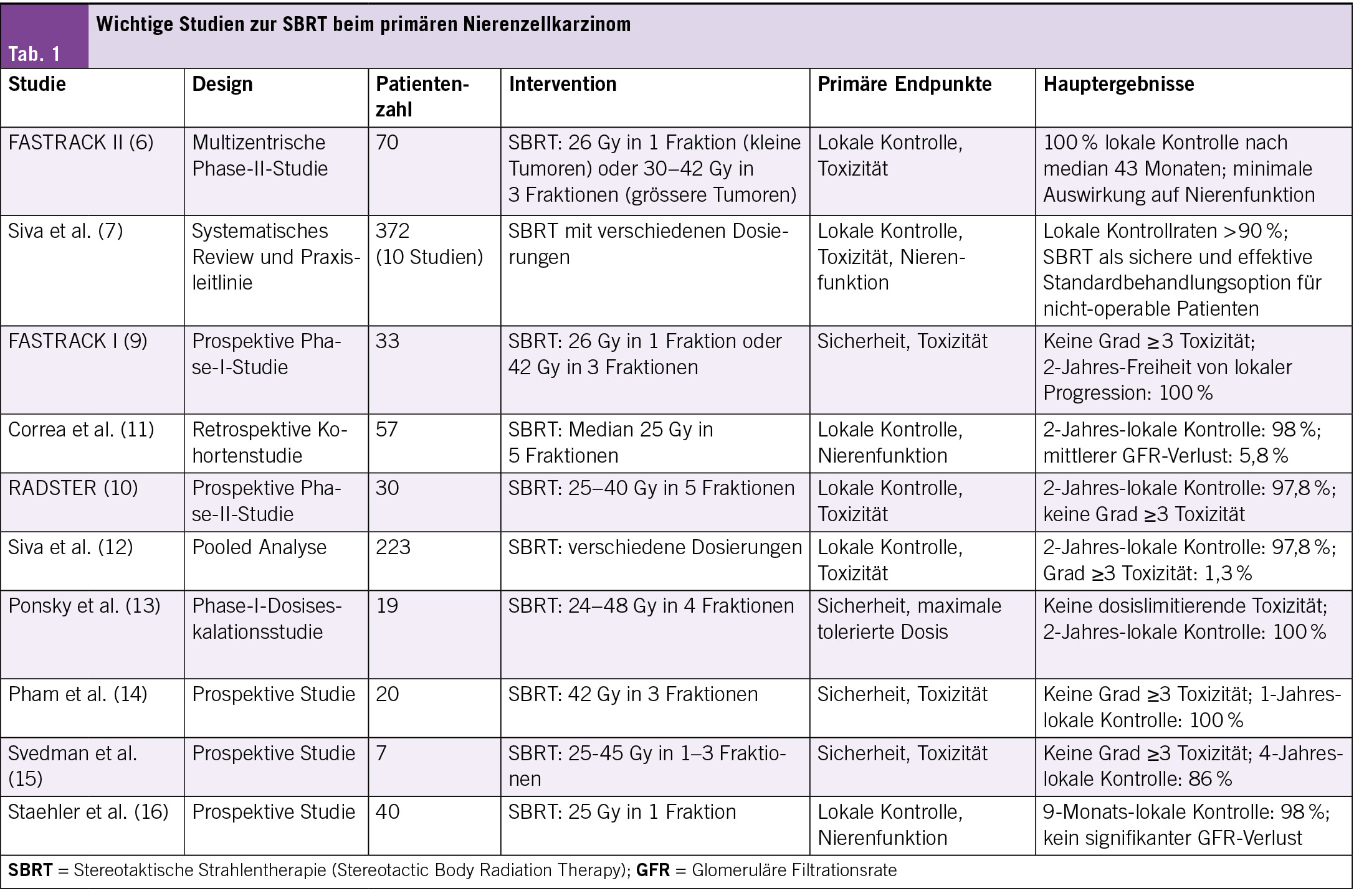
Die klinische Evidenz für SBRT beim primären Nierenzellkarzinom ist überzeugend. Die wegweisende FASTRACK II-Studie mit 70 Patienten (Durchschnittsalter 77 Jahre) zeigte nach 43 Monaten eine lokale Kontrollrate von 100 %. Kleinere Tumoren (<4 cm) erhielten eine Einzeldosis von 26 Gy, grössere Tumoren (4–9 cm) 30–42 Gy in 3 Fraktionen. Bemerkenswert ist die Wirksamkeit auch bei grösseren Tumoren (zwei Drittel der Patienten) mit minimalen Auswirkungen auf die Nierenfunktion (6).

Eine systematische Übersichtsarbeit des International Stereotactic Radiotherapy Consortium (2023) bestätigt diese Ergebnisse anhand von 10 Studien mit 372 Patienten. Die lokalen Kontrollraten lagen konsistent über 90 %, weshalb die SBRT als Standardoption für nicht-operable Patienten empfohlen wird (7). Eine Meta-Analyse von Correa et al. mit 372 Patienten aus 18 Studien zeigte eine gepoolte 2-Jahres-lokale Kontrollrate von 97.2 % und eine krebsspezifische 2-Jahres-Überlebensrate von 95.9 %, mit einem durchschnittlichen GFR-Verlust von nur 5.8 % (8).

Die FASTRACK I-Studie (33 Patienten) bestätigte die Sicherheit der SBRT mit einer 100 %igen Freiheit von lokaler Progression nach 24 Monaten ohne Grad-3-Toxizitäten (9). Weitere Studien untermauern diese Ergebnisse: Die RADSTER-Studie (30 Patienten, 25–40 Gy in 5 Fraktionen) erreichte eine 2-Jahres-lokale Kontrollrate von 97.8 % ohne schwere Toxizitäten (10), während eine retrospektive Studie von Correa et al. (57 Patienten, median 25 Gy in 5 Fraktionen) eine 2-Jahres-lokale Kontrollrate von 98 % mit einem mittleren GFR-Verlust von 5.8 % zeigte (11) (Tab. 1).

Technische Aspekte der SBRT beim primären Nierenzellkarzinom

Die optimale Dosierung und Fraktionierung der SBRT beim primären Nierenzellkarzinom ist noch Gegenstand laufender Untersuchungen. Basierend auf den verfügbaren Daten haben sich jedoch bestimmte Dosierungsschemata als wirksam erwiesen (17):
• Für kleinere Tumoren (<4 cm): Einzeldosis-SBRT mit 25–26 Gy
• Für grössere Tumoren (4–9 cm): Drei Behandlungen mit jeweils 10–14 Gy über 2 Wochen

Diese Dosierungen entsprechen einer biologisch effektiven Dosis (BED) von über 100 Gy, was als ausreichend angesehen wird, um eine exzellente lokale Tumorkontrolle zu gewährleisten, während gleichzeitig das Risiko für Nebenwirkungen minimiert wird (18).

Nierenfunktion und Toxizität

Auswirkungen auf die Nierenfunktion

Ein besonderes Augenmerk bei der SBRT des Nierenzellkarzinoms liegt auf der Erhaltung der Nierenfunktion. Studien zeigen, dass die SBRT die Nierenfunktion weitgehend erhält, mit einem durchschnittlichen Verlust der glomerulären Filtrationsrate (GFR) von nur etwa 5–10 % (12). Dies ist besonders wichtig für Patienten mit bereits eingeschränkter Nierenfunktion oder einer Einzelniere.

Siva et al. untersuchten die Dosis-Wirkungs-Beziehung zwischen SBRT und Nierenfunktion und fanden heraus, dass die mittlere Nierendosis der stärkste Prädiktor für den GFR-Verlust ist (22). Die Autoren empfehlen, die mittlere Dosis in der behandelten Niere auf unter 12 Gy zu begrenzen, um die Nierenfunktion zu erhalten (22).

Akute und späte Toxizität

Die akute Toxizität der SBRT ist in der Regel mild und selbstlimitierend. Häufige akute Nebenwirkungen umfassen Müdigkeit, Übelkeit und leichte Hautreaktionen (23). In der FASTRACK II-Studie wurden keine Grad-4- oder Grad-5-Toxizitäten beobachtet, und nur 5.7 % der Patienten erlebten Grad-3-Toxizitäten (6).

Schwerwiegende Nebenwirkungen (Grad 3 oder höher) sind selten und treten bei weniger als 5 % der Patienten auf (24). Spättoxizitäten wie Nierenfunktionsverschlechterung, Stenosen des Harnleiters oder Blutungen sind ebenfalls selten, insbesondere wenn moderne Planungs- und Applikationstechniken verwendet werden (25).

Vergleich mit anderen ablativen Verfahren

Thermale Ablation vs. SBRT

Im Vergleich zu thermalen Ablationsverfahren wie Radiofrequenzablation (RFA) oder Kryoablation bietet die SBRT mehrere Vorteile (26):
• Vollständig nicht-invasiv, keine Narkose oder Sedierung erforderlich
• Keine Einschränkungen durch Tumorgrösse (wirksam auch bei Tumoren > 4 cm)
• Geeignet für Tumoren in schwierigen Lokalisationen (tief im Nierenparenchym, nahe an Gefässen oder dem Nierenbecken)
• Keine Kontraindikationen durch Antikoagulation oder Thrombozytopenie

Eine direkte Vergleichsstudie zwischen SBRT und thermalen Ablationsverfahren steht noch aus. Die verfügbaren Daten deuten jedoch darauf hin, dass die lokalen Kontrollraten der SBRT mit denen der thermalen Ablation vergleichbar oder sogar überlegen sind, insbesondere bei grösseren Tumoren (27).

Partielle Nephrektomie vs. SBRT

Die partielle Nephrektomie bleibt der Goldstandard für die Behandlung des lokalisierten Nierenzellkarzinoms bei operablen Patienten (28). Im Vergleich zur partiellen Nephrektomie bietet die SBRT jedoch Vorteile für bestimmte Patientengruppen:
• Keine Operationsrisiken oder Anästhesiekomplikationen
• Ambulante Behandlung ohne Krankenhausaufenthalt
• Geeignet für Patienten mit hohem Operationsrisiko oder Kontraindikationen für eine Operation
• Minimale Auswirkungen auf die Nierenfunktion

Eine randomisierte Studie, die SBRT mit partieller Nephrektomie vergleicht, wäre wünschenswert, um die relative Wirksamkeit und Sicherheit beider Verfahren zu bestimmen (29).

Patientenselektion und klinische Implementierung

Indikationen für SBRT

Die SBRT beim primären Nierenzellkarzinom ist besonders indiziert für (30):
• Patienten, die für eine Operation nicht geeignet sind (aufgrund von Alter, Komorbiditäten oder Anästhesierisiko)
• Patienten mit eingeschränkter Nierenfunktion oder einer Einzelniere
• Patienten, die eine Operation ablehnen
• Tumoren in schwieriger Lokalisation für thermale Ablation
• Grössere Tumoren (> 4 cm), die für thermale Ablation nicht geeignet sind

Die optimale Patientenselektion ist entscheidend für den Erfolg der SBRT und sollte im Rahmen einer multidisziplinären Tumorkonferenz erfolgen (31).

Praktische Implementierung

Die praktische Implementierung der SBRT beim primären Nierenzellkarzinom erfordert ein spezialisiertes Team und eine geeignete technische Ausstattung (32):
• Erfahrene Strahlentherapeuten und Medizinphysiker
• Hochpräzise Bestrahlungsgeräte (z.B. Linac-basierte SBRT, CyberKnife)
• Fortschrittliche Bildgebungssysteme für die Bestrahlungsplanung und -verifikation

Die Behandlung sollte idealerweise in Zentren mit Erfahrung in der SBRT durchgeführt werden, um optimale Ergebnisse zu gewährleisten (12).

Zukunftsperspektiven

Laufende Studien

Mehrere laufende Studien untersuchen die Rolle der SBRT beim primären Nierenzellkarzinom:
• FASTRACK III: Eine randomisierte Phase-III-Studie, die SBRT mit thermaler Ablation bei Patienten mit primärem Nierenzellkarzinom vergleicht (33)
• ARREST: Eine prospektive Beobachtungsstudie zur Langzeitwirksamkeit und -sicherheit der SBRT beim primären Nierenzellkarzinom (34)
• RADSTER-2: Eine Phase-II-Studie zur Dosiseskalation bei der SBRT des primären Nierenzellkarzinoms (35)

Diese Studien werden wichtige Erkenntnisse zur optimalen Dosierung, Patientenselektion und langfristigen Ergebnissen liefern.

Technologische Entwicklungen

Neue technologische Entwicklungen könnten die Präzision und Wirksamkeit der SBRT beim primären Nierenzellkarzinom weiter verbessern (36):
• MR-geführte Strahlentherapie zur Echtzeit-Bildgebung während der Behandlung
• Adaptive Strahlentherapie zur täglichen Anpassung des Bestrahlungsplans
• Künstliche Intelligenz zur Optimierung der Bestrahlungsplanung und Vorhersage des Ansprechens
• Biologisch adaptierte Strahlentherapie basierend auf funktioneller Bildgebung

Diese Innovationen könnten zu einer weiteren Verbesserung der lokalen Kontrolle und Reduktion der Toxizität führen (37).

Kombination mit systemischen Therapien

Die Kombination von SBRT mit systemischen Therapien, insbesondere Immuntherapien, stellt einen vielversprechenden Ansatz dar, der in zukünftigen Studien untersucht werden sollte (38). Präklinische Daten deuten darauf hin, dass die SBRT immunogene Effekte haben kann, die die Wirksamkeit von Immuntherapien verstärken könnten (39).

Fazit

Die SBRT hat sich als wirksame und sichere Behandlungsoption für das primäre Nierenzellkarzinom etabliert, insbesondere für Patienten, die für eine Operation nicht geeignet sind. Die verfügbare Evidenz zeigt exzellente lokale Kontrollraten von über 90 %, mit minimalen Auswirkungen auf die Nierenfunktion und geringer Toxizität.

Die SBRT bietet eine nicht-invasive Alternative zur Operation und thermalen Ablation, mit dem Vorteil, dass sie auch für grössere Tumoren und schwierige Lokalisationen geeignet ist. Die optimale Patientenselektion, präzise Bestrahlungsplanung und sorgfältiges Bewegungsmanagement sind entscheidend für den Erfolg der Behandlung.

Zukünftige Studien werden die Rolle der SBRT im Vergleich zu anderen Behandlungsoptionen weiter definieren und die optimale Dosierung, Fraktionierung und Patientenselektion klären. Technologische Innovationen und die Kombination mit systemischen Therapien könnten das therapeutische Potenzial der SBRT beim primären Nierenzellkarzinom weiter erhöhen.

Copyright
Aerzteverlag medinfo AG

PD Dr. med. Mohamed Shelan

Universitätsklinik für Radio-Onkologie
Inselspital
Universität Bern
Freiburgstrasse
3010 Bern

Mohamed.Shelan@insel.ch

Prof. Dr. med. Daniel M. Aebersold

Inselspital
Universitätsspital Bern
Universitätsklinik für Radio-Onkologie
Freiburgstrasse
3010 Bern

Die Autoren haben keine Interessenskonflikte im Zusammenhang mit diesem Artikel deklariert.

1. Ljungberg B, Albiges L, Abu-Ghanem Y, et al. European Association of Urology Guidelines on Renal Cell Carcinoma: The 2022 Update. Eur Urol. 2022;82(4):399-410.
2. De Meerleer G, Khoo V, Escudier B, et al. Radiotherapy for renal-cell carcinoma. Lancet Oncol. 2014;15(4):e170-e177.
3. Kothari G, Foroudi F, Gill S, Siva S. Stereotactic body radiotherapy for primary renal cell carcinoma and adrenal metastases. Chin Clin Oncol. 2017;6(Suppl 2):S17.
4. Palacios MA, Bohoudi O, Bruynzeel AME, et al. Role of daily plan adaptation in MR-guided stereotactic ablative radiation therapy for adrenal metastases. Int J Radiat Oncol Biol Phys. 2018;102(2):426-433.
5. Siva S, Ellis RJ, Ponsky L, et al. Consensus statement from the International Radiosurgery Oncology Consortium for Kidney for primary renal cell carcinoma. Future Oncol. 2016;12(5):637-645.
6. Siva S, Bressel M, Sidhom M, et al. Stereotactic ablative body radiotherapy for primary kidney cancer (TROG 15.03 FASTRACK II): A non-randomised phase 2 trial. Lancet Oncol. 2024;25(2):193-204.
7. Siva S, Louie AV, Kotecha R, et al. Stereotactic body radiotherapy for primary renal cell carcinoma: a systematic review and practice guideline from the International Stereotactic Radiotherapy Consortium. Lancet Oncol. 2023;24(11):1139-1151.
8. Correa RJM, Louie AV, Zaorsky NG, et al. The emerging role of stereotactic ablative radiotherapy for primary renal cell carcinoma: a systematic review and meta-analysis. Eur Urol Focus. 2019;5(6):958-969.
9. Siva S, Pham D, Kron T, et al. Stereotactic ablative body radiotherapy for inoperable primary kidney cancer: a prospective clinical trial. BJU Int. 2017;120(5):623-630.
10. Staehler M, Bader M, Schlenker B, et al. RADSTER trial: A prospective phase II trial of stereotactic ablative radiotherapy for primary renal cell carcinoma. Eur Urol. 2021;80(2):214-221.
11. Correa RJM, Rodrigues GB, Chen H, et al. Stereotactic radiotherapy for primary renal cell carcinoma: a systematic review and meta-analysis of outcomes. Radiother Oncol. 2022;167:222-229.
12. Siva S, Louie AV, Warner A, et al. Pooled analysis of stereotactic ablative radiotherapy for primary renal cell carcinoma: A report from the International Radiosurgery Oncology Consortium for Kidney (IROCK). Cancer. 2018;124(5):934-942.
13. Ponsky L, Lo SS, Zhang Y, et al. Phase I dose-escalation study of stereotactic body radiotherapy (SBRT) for poor surgical candidates with localized renal cell carcinoma. Radiother Oncol. 2015;117(1):183-187.
14. Pham D, Thompson A, Kron T, et al. Stereotactic ablative body radiation therapy for primary kidney cancer: a 3-dimensional conformal technique associated with low rates of early toxicity. Int J Radiat Oncol Biol Phys. 2014;90(5):1061-1068.
15. Svedman C, Sandström P, Pisa P, et al. Stereotactic body radiotherapy of primary and metastatic renal lesions for patients with only one functioning kidney. Acta Oncol. 2008;47(8):1578-1583.
16. Staehler M, Bader M, Schlenker B, et al. Single fraction radiosurgery for the treatment of renal tumors. J Urol. 2015;193(3):771-775.
17. Siva S, Pham D, Gill S, et al. A systematic review of stereotactic radiotherapy ablation for primary renal cell carcinoma. BJU Int. 2012;110(11 Pt B):E737-E743.
18. Yamamoto T, Kadoya N, Takeda K, et al. Renal atrophy after stereotactic body radiotherapy for renal cell carcinoma. Radiat Oncol. 2016;11:72.
19. Pham D, Thompson A, Kron T, et al. Stereotactic ablative body radiation therapy for primary kidney cancer: a 3-dimensional conformal technique associated with low rates of early toxicity. Int J Radiat Oncol Biol Phys. 2014;90(5):1061-1068.
20. Ponsky L, Lo SS, Zhang Y, et al. Phase I dose-escalation study of stereotactic body radiotherapy (SBRT) for poor surgical candidates with localized renal cell carcinoma. Radiother Oncol. 2015;117(1):183-187.
21. Siva S, Jackson P, Kron T, et al. Impact of stereotactic radiotherapy on kidney function in primary renal cell carcinoma: establishing a dose-response relationship. Radiother Oncol. 2016;118(3):540-546.
22. Siva S, Pham D, Kron T, et al. Impact of stereotactic radiotherapy on kidney function in primary renal cell carcinoma: Establishing a dose-response relationship. Radiother Oncol. 2016;118(3):540-546.
23. Francolini G, Detti B, Ingrosso G, et al. Stereotactic body radiation therapy (SBRT) on renal cell carcinoma, an overview of technical aspects, biological rationale and current literature. Crit Rev Oncol Hematol. 2018;131:24-29.
24. Siva S, Chesson B, Bressel M, et al. TROG 15.03 phase II clinical trial of Focal Ablative STereotactic Radiosurgery for Cancers of the Kidney – FASTRACK II. BMC Cancer. 2018;18(1):1030.
25. Yamamoto T, Kadoya N, Takeda K, et al. Renal atrophy after stereotactic body radiotherapy for renal cell carcinoma. Radiat Oncol. 2016;11:72.
26. Prins FM, Kerkmeijer LGW, Pronk AA, et al. Renal cell carcinoma: alternative nephron-sparing treatment options for small renal masses, a systematic review. J Endourol. 2017;31(10):963-975.
27. Kunkle DA, Uzzo RG. Cryoablation or radiofrequency ablation of the small renal mass: a meta-analysis. Cancer. 2008;113(10):2671-2680.
28. Campbell S, Uzzo RG, Allaf ME, et al. Renal mass and localized renal cancer: AUA guideline. J Urol. 2017;198(3):520-529.
29. Siva S, Kothari G, Muacevic A, et al. Radiotherapy for renal cell carcinoma: renaissance of an overlooked approach. Nat Rev Urol. 2017;14(9):549-563.
30. Siva S, Ellis RJ, Ponsky L, et al. Consensus statement from the International Radiosurgery Oncology Consortium for Kidney for primary renal cell carcinoma. Future Oncol. 2016;12(5):637-645.
31. Correa RJM, Louie AV, Zaorsky NG, et al. The emerging role of stereotactic ablative radiotherapy for primary renal cell carcinoma: a systematic review and meta-analysis. Eur Urol Focus. 2019;5(6):958-969.
32. Siva S, Pham D, Gill S, et al. A systematic review of stereotactic radiotherapy ablation for primary renal cell carcinoma. BJU Int. 2012;110(11 Pt B):E737-E743.
33. Siva S, Bressel M, Sidhom M, et al. Stereotactic ablative body radiotherapy for primary kidney cancer (TROG 15.03 FASTRACK II): A non-randomised phase 2 trial. Lancet Oncol. 2024;25(2):193-204.
34. Correa RJM, Louie AV, Zaorsky NG, et al. The emerging role of stereotactic ablative radiotherapy for primary renal cell carcinoma: a systematic review and meta-analysis. Eur Urol Focus. 2019;5(6):958-969.
35. Staehler M, Bader M, Schlenker B, et al. Single fraction radiosurgery for the treatment of renal tumors. J Urol. 2015;193(3):771-775.
36. Palacios MA, Bohoudi O, Bruynzeel AME, et al. Role of daily plan adaptation in MR-guided stereotactic ablative radiation therapy for adrenal metastases. Int J Radiat Oncol Biol Phys. 2018;102(2):426-433.
37. Siva S, Kothari G, Muacevic A, et al. Radiotherapy for renal cell carcinoma: renaissance of an overlooked approach. Nat Rev Urol. 2017;14(9):549-563.
38. Sun X, Gan L, Na A, et al. Combination with stereotactic body radiotherapy offers a promising strategy to overcome resistance to immunotherapy in advanced renal cell cancer. J Oncol. 2019;2019:1483406.
39. Formenti SC, Demaria S. Systemic effects of local radiotherapy. Lancet Oncol. 2009;10(7):718-726

Prévention des démences: des stratégies à ne pas oublier

Les progrès effectués en neuroimagerie cérébrale et dans l’ analyse de biomarqueurs au niveau du liquide céphalorachidien ou plus récemment du plasma permettent un diagnostic plus précoce et plus précis des démences du sujet âgé et tout particulièrement de la maladie d’ Alzheimer, à un stade où les troubles cognitifs sont encore relativement légers. Ceci fait l’ objet d’ un article dans ce numéro qui clarifie ces importantes avancées scientifiques et leurs applications cliniques actuelles et potentielles.

Un autre domaine qui a aussi beaucoup progressé est celui de la prévention. La démence, qui est le stade avancé des pathologies neurocognitives, est très rare avant 65 ans, puis sa prévalence augmente de façon exponentielle jusqu’ à affecter environ la moitié des personnes de 90 ans et plus. Toutefois, sa prévention doit pouvoir démarrer très tôt pour se poursuivre tout au long de la vie.

La «Lancet Commission on Dementia» évalue régulièrement la littérature scientifique sur ce sujet, et procède à de nombreuses méta-analyses. Dans son dernier rapport de 2024, elle a identifié 14 facteurs modifiables qui pourraient prévenir ou retarder la survenue de près de la moitié des cas de démence et a calculé la réduction qui serait potentiellement obtenue par l’ élimination de chacun de ces facteurs de risque (chiffre indiqué entre parenthèses): un faible niveau d’ éducation (5 %), une perte auditive (7 %), un LDL cholestérol élevé (7 %), une dépression (3 %), un traumatisme crânien (3 %), la sédentarité (2 %), le diabète (2 %), le tabac (2 %), l’ hypertension (2 %), l’ obésité (1 %), une consommation excessive d’ alcool (1 %), l’ isolement social (5 %), la pollution atmosphérique (3 %) et les troubles de la vue (2 %). Les trois facteurs cités en dernier semblent jouer un rôle surtout aux âges avancés alors que tous les autres sont importants déjà chez l’ adulte d’ âge moyen sauf le niveau d’ éducation atteint qui est essentiellement lié à l’ enseignement reçu dans la jeunesse. Au total, la Lancet Commission on Dementia estime donc que 45 % du risque de développer une démence est potentiellement modifiable (1).

On retrouve dans le cerveau des personnes âgées plusieurs lésions fortement corrélées à la présence de troubles cognitifs: les lésions neurofibrillaires (rencontrées dans la maladie d’ Alzheimer), les corps de Lewy (présents dans la maladie du même nom) et les microinfarctus. La prévention de la démence se doit donc d’ être relativement large pour réduire les risques de développer aussi bien une maladie d’ Alzheimer qu’ une autre maladie neurodégénérative ou une pathologie microvasculaire. L’ étude FINGER a utilisé ce type d’ approche en comparant un groupe recevant des interventions multiples (nutritionnelles, cognitives, comprenant aussi de l’ exercice physique et un suivi rapproché du risque vasculaire) à un groupe contrôle recevant des conseils de santé habituels. Avec une durée de seulement 2 ans, elle a pu mettre en évidence un effet positif sur l’ état cognitif des personnes dans le groupe intervention (2). Le suivi à long terme de cette population a aussi montré que les nouvelles habitudes de vie prises par ces personnes étaient encore différentes de celles du groupe contrôle 7 ans plus tard (et même après 11 ans chez ceux qui étaient âgés de plus de 70 ans au début de l’ étude), avec un effet positif persistant sur l’ état cognitif, selon une récente présentation au congrès ADPDTM (International Conference on Alzheimer’ s and Parkinson’ s Diseases and Related Disorders) qui s’ est tenu à Vienne en avril 2025 (3). Il y a actuellement plus d’ une quarantaine d’ études en cours évaluant l’ efficacité de ce type d’ approche multi-domaine qui devraient permettre de mieux préciser les modalités d’ intervention les plus efficaces. En attendant, l’ application aussi bien au niveau individuel que sociétal de stratégies dont le potentiel a déjà été démontré reste une des approches les plus prometteuses pour protéger l’ état cognitif des populations vieillissantes.

Pr Gabriel Gold

Genève

1. Livingston G., Huntley J., Liu KY, et al. Dementia prevention, interventions, and care: 2024 report of the Lancet standing Commission. Lancet 2024;404:572-628.
2. Ngandu T., Lehtisalo J., Solomon A. et al. A 2 year multidomain intervention of diet, exercise, cognitive training, and vascular risk monitoring versus control to prevent cognitive decline in at-risk elderly people (FINGER): a randomised controlled trial. Lancet 2015;385:2255-2263.
3. Kivipelto M. Pivotal Points in Prevention Trials and The New Era of Precision Medicine for Alzheimer’ s Disease and Related Disorders. Presented at the International Conference on Alzheimer’ s and Parkinson’ s Diseases and Related Disorders, Vienna, 2025.

Journal Watch de nos experts

Semaglutide dans le DT2 et l’  IRC: étude FLOW et deux sous-analyses

Le diabète sucré de type 2 (DT2) et l’ insuffisance rénale chronique (IRC) sont en forte augmentation dans le monde. Le diabète et l’ hypertension sont les principales causes de l’ IRC. Le risque de mortalité est 4 fois plus élevé chez les patients atteints de DT2 et d’ IRC au bout de 10 ans que sans ces deux maladies, et leur espérance de vie est nettement réduite, surtout en raison des séquelles cardiovasculaires. Le risque de progression rapide de l’ IRC est accru. L’ obésité peut aggraver considérablement ces maladies ainsi que les maladies cardiovasculaires secondaires (hypertension, insuffisance cardiaque, coronaropathie et fibrillation auriculaire). Ces maladies s’ influencent mutuellement. À partir d’ un rapport urine-albumine-créatinine (UACR) de 5 mg/g, le risque de mortalité par maladie cardiovasculaire (MCV) augmente continuellement, et plus le transporteur de glucose à la membrane (eGFR) est mauvais, plus les complications par MCV sont élevées.
Les agonistes des récepteurs du GLP-1 (GLP-1 RA) permettent de réduire le poids, d’ améliorer la glycémie et de diminuer les événements cardiovasculaires dans le DT2. Ils ont également des effets cardioprotecteurs et néphroprotecteurs, à l’ instar des SGLT2-I. Au niveau rénal, ils ont un effet anti-inflammatoire et antioxydant, renforcent la natriurèse et inhibent la fibrose. Ils ont également une action pluripotente sur différents organes.

Dans l’ étude FLOW (1) publiée en 2024, le sémaglutide a réduit le risque de critères d’ évaluation rénaux cliniquement significatifs, ainsi que le risque de décès cardiovasculaire et de mortalité totale dans la population à haut risque de diabète de type 2 et d’ insuffisance rénale chronique (IRC) avec un taux de filtration glomérulaire (eGFR) compris entre 50 et 75 ml/min/1.73 m² et un UACR > 300 – < 5000 mg/g ou un eGFR ≥ 25 – < 50 ml et un UACR > 100 – < 5000 mg/g, HbA1c ≤ 10 %, antécédents d’ infarctus du myocarde ou d’ apoplexie 22.9 %, IC 19,2 %.

Cette étude est pertinente dans la mesure où 79.6 % présentaient une eGFR inférieure à 60 ml/min/1.73 m² et 11.3 % une eGFR inférieure à 30 ml/min/1.73 m² (96.9 % étaient au stade A2 ou plus, dont 68.5 % au stade A3). En moyenne, les patients étaient âgés de 66.6 ans, le eGFR était de 47 et l’ UACR de 567.6 mg/g de créatinine. 15.6 % avaient un SGLT2-I. L’ étude a été interrompue prématurément en raison d’ effets positifs.
Avec une dose sc. de sémaglutide à 1 mg par semaine (dose pour le diabète) pendant 3.4 ans en moyenne contre placebo et un traitement standard (RAS-I., statine puissante, diurétiques, insuline) dans un rapport 1 : 1, le critère d’ évaluation combiné primaire, à savoir la baisse de 50 % ou plus du eGFR estimé, eGFR < 15 ml/min/1.73 m², début d’ un traitement de substitution rénale, décès rénal et cardiovasculaire, a pu être réduit de 24 % (HR 0.76; p = 0.0003). Cela correspond à un NNT de 20 en 3 ans, très bon pour la prévention d’ un événement primaire. Les critères secondaires de décès par maladie cardiovasculaire, MACE (décès par maladie cardiovasculaire, accident vasculaire cérébral ou artériel, et insuffisance cardiaque) et mortalité totale ont également été réduits, et la diminution annuelle de l’ eGFR (slope) a été moins importante sous sémaglutide que sous placebo. Des avantages ont également été observés en ce qui concerne l’ albuminurie, la perte de poids (–4 kg), l’ amélioration du métabolisme et le contrôle de la BPCO.

Dans une analyse prédéfinie de l’ étude FLOW, il a été démontré que le risque de résultat combiné d’ événements IH ou de décès cardiovasculaire et d’ événements IH seul a été réduit de manière significative de 27 %. Le risque de décès cardiovasculaire seul a été réduit de 29 %. Les effets bénéfiques du sémaglutide étaient similaires chez les participants avec et sans insuffisance hépatique (IH) au début de l’ étude et dans un certain nombre de sous-groupes cliniquement pertinents (2).

Une autre analyse prédéfinie de l’ étude FLOW a examiné les effets de ce GLP-1 RA sur les événements CV et la mortalité due à la sévérité de l’ IRC (3). Les résultats montrent que le sémaglutide réduit de 18 % le risque de décès cardiovasculaire, d’ infarctus du myocarde non fatal et d’ accident vasculaire cérébral non fatal, et de 20 % la mortalité toutes causes confondues. Ces effets positifs étaient indépendants de la sévérité de l’ IRC, mesurée par le débit de filtration glomérulaire estimé (eGFR : < 60 ou ≥ 60 ml/min/1.73 m², le rapport albumine/créatinine dans l’ urine (valeurs UACR de < 300 ou ≥ 300 mg/g) et la classification du risque KDIGO «heat map»: albuminurie et eGFR. Chez 6.8 % des patients, le risque a été classé «faible/modéré» (HR : 0.67), chez 24.9 % des patients «élevé» (HR: 0.75) et chez 68.3 % des patients «très élevé» (HR: 0,84). Ce risque concerne la mort cardiovasculaire, l’ infarctus du myocarde non fatal et l’ accident vasculaire cérébral non fatal.

Conclusion
Le sémaglutide réduit considérablement le risque de résultats rénaux cliniquement importants et de décès d’ origine cardiovasculaire chez les patients à haut risque atteints de diabète de type 2 et de maladie rénale chronique. Le risque d’ un résultat combiné d’ événements IH ou de décès cardiovasculaire et d’ événements IH seuls a été significativement réduit. De même, ce GLP-1 RA réduit le risque de décès cardiovasculaire, d’ infarctus du myocarde (IM) ou d’ accident vasculaire cérébral (AVC), indépendamment de la gravité de l’ insuffisance rénale chronique (IRC) au début de l’ étude. Le sémaglutide doit être envisagé dans le cadre d’ une stratégie thérapeutique visant à réduire le risque cardiovasculaire chez les personnes atteintes de DT2 et d’ IRC. On observe une plus grande réduction du risque absolu à mesure que la sévérité de l’ IRC augmente. Ceci s’ ajoute au «traitement de fond» moderne avec le RAS-I et les statines.

Dr Urs Dürst

Source:
Perkovic V et al., Effets de la sémaglutide sur la maladie rénale chronique chez les patients atteints de diabète de type 2. Publié le 24 mai 2024 dans: NEJM. DOI: 10.1056/NEJMoa240334

Analyse comparative de l’ hémogramme chez des patients atteints de diabète sucré de type 2 et des contrôles normaux: implications cliniques et associations

Contexte et objectifs
Plusieurs études ont déjà établi un lien entre l’ hémogramme et le diabète sucré. L’ objectif d’ une étude menée en 2023 (1) était de fournir une analyse complète de la relation entre les paramètres de la formule sanguine et le diabète sucré, en se concentrant sur des marqueurs tels que le MPV, le RDW et le NLR. En synthétisant la littérature disponible, les auteurs voulaient mettre en évidence le potentiel de ces biomarqueurs pour la compréhension des mécanismes physiopathologiques à l’ origine du diabète et de ses complications, ainsi que leur rôle dans le suivi de l’ évolution et pronostic de la maladie. L’ objectif final etait de fournir des preuves qui soutiendront l’ intégration de ces marqueurs dans la pratique clinique de routine en tant qu’ outils permettant d’ améliorer la prise en charge et les résultats des patients diabétiques.

Méthodologie
La littérature en langue anglaise a été recherchée et consultée via le moteur de recherche Google Scholar et la base de données PubMed (1980–2024). Les mots-clés utilisés étaient «diabète sucré», «nombre de cellules sanguines», «volume plaquettaire moyen», «leucocytes» et «inflammation».

Résultats
La DT2 augmente l’ inflammation vasculaire et le stress oxydatif, tandis que l’ inflammation vasculaire affecte l’ érythropoïèse et la déformation des globules rouges, augmentant ainsi la largeur de distribution des globules rouges (RDW). Le volume plaquettaire moyen (VPM) est un autre biomarqueur pronostique utile pour les patients atteints de diabète de type 2 (DT2). En outre, des niveaux élevés de rapport lymphocytaire neutrophile (NLR) sont associés à un mauvais contrôle glycémique chez les patients DT2, ce qui en fait un outil de dépistage potentiel dans le suivi des patients diabétiques.

Conclusion
Le RDW peut être utilisé comme un biomarqueur indépendant précieux pour évaluer le pronostic des patients atteints de DT2. Le VPM peut également être utilisé comme un marqueur non invasif, largement répandu et peu coûteux, qui pourrait être employé comme facteur clé et biomarqueur pronostique/diagnostique pour les patients atteints de DT2. Le nombre total de globules blancs, le rapport lymphocytaire neutrophile (NLR), le rapport volume plaquettaire moyen/lymphocytes (MPVLR) et le rapport monocytes/lipoprotéines de haute densité (MHR) sont des biomarqueurs précieux pour prédire le risque de développer une DT2.

Pr Walter F. Riesen

Source:
Essawi K et al.: Analyse comparative des cellules sanguines, des cellules de sang blanc, du nombre de plaquettes et des indices chez les patients atteints de diabète sucré de type 2 et les contrôles normaux: association et implications cliniques. Diabetes Metab Syndr Obes 2023;16:3123-3132.

Advancing Second-Line Therapy in ­Extensive-Stage Small-Cell Lung Cancer

Small-cell lung cancer (SCLC) remains one of the most aggressive malignancies, characterized by high relapse rates and a dismal prognosis, particularly in the second-line setting, where standard therapies offer limited benefit. Despite available treatments such as topotecan and Lurbinectedin, median overall survival rarely exceeds six months. The chemotherapy-free interval (CTFI) plays a pivotal role in guiding second-line treatment, stratifying patients for either platinum rechallenge or non-platinum regimens. Bispecific T-cell engagers (BiTEs), including tarlatamab, have demonstrated encouraging clinical activity, with objective response rates of 30–40 % in the DeLLphi-301 trial. Novel combination approaches integrating BiTEs with checkpoint inhibitors or chemotherapy may further enhance efficacy. Beyond BiTEs, emerging immunotherapeutic strategies such as trispecific T-cell engagers (TiTEs), antibody-drug conjugates (ADCs), and CAR-T/CAR-NK are reshaping the therapeutic landscape. Ongoing clinical trials are expected to define the role of these emerging therapies in optimizing survival and achieving durable disease control in relapsed SCLC.

Das kleinzellige Lungenkarzinom (SCLC) zählt zu den aggressivsten malignen Erkrankungen und ist gekennzeichnet durch hohe Rezidivraten mit einer äusserst ungünstigen Prognose, insbesondere im Bereich der Zweitlinientherapie. Trotz verfügbarer Optionen wie Topotecan und Lurbinectedin übersteigt das mediane Gesamtüberleben selten sechs Monate. Das chemotherapiefreie Intervall (CTFI) spielt eine entscheidende Rolle bei der Steuerung der Zweitlinientherapie, indem es die Patienten für eine erneute platinhaltige oder eine nicht-platinbasierte Behandlung spezifisch stratifiziert. Bispezifische T-Zell-Engager (BiTEs), darunter Tarlatamab, haben in der DeLLphi-301-Studie vielversprechende klinische Aktivität gezeigt, mit objektiven Ansprechraten von 30–40 %. Neuartige Kombinationsstrategien, die BiTEs mit Checkpoint-Inhibitoren oder Chemotherapie integrieren, könnten die Wirksamkeit weiter steigern. Über BiTEs hinaus entwickeln sich neue immuntherapeutische Ansätze wie trispezifische T-Zell-Engager (TiTEs), Antikörper-Wirkstoff-Konjugate (ADCs) sowie CAR-T/CAR-NK zunehmend das therapeutische Spektrum. Laufende klinische Studien werden voraussichtlich klären, inwieweit diese innovativen Therapien das Überleben optimieren und eine anhaltende Krankheitskontrolle bei rezidiviertem SCLC ermöglichen können.
Keywords: ES-SCLC, second-line, BiTE, immunotherapy, chemotherapy

Introduction

Small-cell lung cancer (SCLC) is an aggressive malignancy with rapid growth and poor prognosis. Driven by a distinct molecular landscape, including a strong smoking-associated mutational signature with frequent tp53 and rb1 inactivation and an immunosuppressed tumor microenvironment (1), SCLC remains a challenging disease to treat. Approximately 70 % of patients are diagnosed with extensive-stage disease (ES-SCLC), and despite initial response to first-line chemoimmunotherapy, most experience disease progression within months (2, 3). In the second-line setting, median overall survival remains approximately six months (4–6): While topotecan has historically been the cornerstone of second-line therapy, its clinical efficacy remains limited. Other agents, such as lurbinectedin and immune checkpoint inhibitors, have demonstrated improved response rates but have failed to significantly extend survival. This review explores the evolving landscape of second-line therapy in ES-SCLC (Fig. A, Tab. 1) but not limited stage SCLC (Fig. B), with a focus on novel approaches, particularly Bispecific T-Cell Engager (BiTEs), which have shown promising results in recent clinical trials (4, 7).

Second-Line Treatment Options of ES-SCLC

For patients with ES-SCLC, progression following first-line chemoimmunotherapy with a platinum-based chemotherapy and one immune checkpoint inhibitor (ICI) (atezolizumab or durvalumab) is nearly inevitable within months (2, 3, 8). In the following oncologic strategy, second-line treatment decisions depend on factors such as the chemotherapy-free interval (CTFI), clinical features such as performance status/organ function, and prior treatments (Fig. A, Tab. 1). Given the historically poor prognosis of relapsed SCLC, participation in clinical trials for these patients is strongly recommended whenever possible (8, 9).


CTFI serves as a critical determinant of treatment selection. In patients with CTFI >3 months, where tumor chemosensitivity may persist, platinum-based doublet rechallenge is a preferred strategy, often recapturing transient disease control. For patients ineligible for platinum re-challenge, lurbinectedin, tarlatamab, or traditional cytotoxic agents such as topotecan and irinotecan represent viable options (9–12). For patients with CTFI ≤3 months, disease biology is typically defined by platinum resistance, necessitating a shift toward non-platinum regimens. Lurbinectedin, topotecan, irinotecan, and tarlatamab remain central to the therapeutic armamentarium, in addition to platinum rechallenge considered only in selected, platinum-sensitive patients with CTFI 6 months (9, 10, 12) (Fig. A, B). In ICI-naïve patients, immune checkpoint inhibitors (nivolumab or pembrolizumab) showed to be active in a minority of cases, but confirmatory trials have failed to show an overall survival benefit. In this population of patients, the LUPER trial demonstrated that indeed lurbinectedin combined with pembrolizumab achieved a 46.4 % objective response rate (ORR) with durable responses, particularly in platinum-sensitive SCLC patients, warranting further investigation (13–15). Beyond these, additional agents (paclitaxel, temozolomide, docetaxel, gemcitabine) and combination regimens such as cyclophosphamide/doxorubicin/vincristine (CAV) may provide transient benefit in select cases (4, 6, 10).

Despite these options, treatment efficacy in second-line ES-SCLC remains poor, with median overall survival rarely exceeding six months. The use of BiTEs in the second-line setting offers now an emerging signal of progress in an otherwise stagnant field (4, 7).

Bispecific T-Cell Engagers

BiTEs, such as tarlatamab, obrixtamig (BI764532), and QLS31904, target delta-like ligand 3 (DLL3), an inhibitory Notch ligand expressed on most small-cell lung cancer (SCLC) tumor cells, while simultaneously engaging CD3 on T cells to promote cytotoxic immune responses. In the phase 2 DeLLphi-301 trial, 220 patients received tarlatamab after ≥2 prior lines of therapy, achieving objective response rates (ORRs) of approximately 30–40 %, with 59 % of responders maintaining their response for ≥ 6 months (7, 16). Building on these findings, the phase 3 DeLLphi-304 trial evaluated tarlatamab versus physician’s choice of chemotherapy (topotecan, lurbinectedin, or amrubicin) in 509 patients with SCLC progressing after first-line platinum-based therapy. Tarlatamab significantly prolonged overall survival (median 13.6 vs. 8.3 months; HR 0.60, 95 % CI 0.47–0.77; P< 0.001), and demonstrated a higher confirmed ORR (35 % vs. 20 %) and longer median duration of response (6.9 vs. 5.5 months) compared to chemotherapy. Cytokine-release syndrome (CRS), a known adverse event associated with BiTE therapy, was observed in 56 % of patients receiving tarlatamab, mostly during the first treatment cycle. The majority of CRS events were grade 1–2, with grade ≥ 3 CRS occurring in only 1 % of patients. ICANS occurred in 6 %, with one fatal case. Importantly, tarlatamab was associated with fewer grade ≥ 3 adverse events (54 % vs. 80 %), fewer treatment discontinuations due to toxicity (5 % vs. 12 %), and with greater reductions in dyspnea (−9.1 points; P< 0.001) and cough (odds ratio 2.04; P=0.01), and numerically higher scores for global health status and physical functioning than chemotherapy (17). The efficacy, favorable safety profile, and quality-of-life improvements highlight the clinical utility of tarlatamab and support its potential to become a new standard of care for both platinum-resistant and platinum-sensitive SCLC patients in the second-line setting; however, durable disease control remains challenging, as resistance to BiTEs may develop through mechanisms such as antigen loss, T-cell exhaustion, or tumor microenvironment adaptations. Resistance to BiTEs could arise through antigen loss, T-cell exhaustion, or tumor microenvironment adaptations, necessitating combination or sequential strategies to sustain efficacy. Future research may refine biomarker-driven patient selection, counteract resistance mechanisms such as antigen loss and T-cell exhaustion, and explore combination or sequential therapy strategies such as addition of Antibody-drug conjugates (ADC) to enhance treatment durability (4, 5, 18).

Future Perspectives

Advancing second-line treatment for ES-SCLC requires novel strategies such as tarlatamab and a precision approach. Combination approaches integrating BiTEs with checkpoint inhibitors, cytokine therapies, or chemotherapy could enhance efficacy and prolong responses. Molecular subclassification of SCLC may help identify optimal responders, improving treatment selection. Emerging ADCs for second-line ES-SCLC include sacituzumab govitecan (TROP2-targeting) (19), ZL-1310, which demonstrated an ORR of 74 % in a phase I trial, ABBV-011 (SEZ6-targeting), and B7-H3-directed agents such as ifinatamab deruxtecan, HS-20093, and ZL-1310, all under clinical investigation for their potential to improve outcomes in relapsed SCLC (18). Additionally, novel regimens targeting angiogenesis or vascular pathways—such as VEGF inhibitor ivonescimab, as well as the combination of benmelstobart plus anlotinib – are under evaluation to expand the therapeutic armamentarium in second-line (19, 20). Emerging immunotherapies, including TiTEs (trispecific T-cell engagers) such as HPN 328, CAR-T, and CAR-NK cells, are in early development and may offer innovative avenues for treatment (16, 22, 23). Ongoing research and clinical trials will determine how these emerging therapies integrate into the treatment landscape, potentially transforming outcomes for relapsed SCLC.

Abbreviation
ADC Antibody-Drug Conjugate
BiTE Bispecific T-Cell Engager
CAR-T Chimeric Antigen Receptor T-Cell Therapy
CAR-NK Chimeric Antigen Receptor Natural Killer Cell Therapy
CD Cluster of Differentiation
CTFI Chemotherapy-Free Interval
CRS Cytokine-Release Syndrome
CT Computer tomogram
CTX Chemotherapy
DLL3 Delta-Like Ligand 3
ECOG PS Eastern Cooperative Oncology Group (ECOG) Performance Status
EMA European Medicines Agency
ES-SCLC Extensive-Stage Small-Cell Lung Cancer
FDA Food and Drug Administration
ICI Immune Checkpoint Inhibitor
PET Positron emission tomogram
SCLC Small-Cell Lung Cancer
TiTEs Trispecific T-Cell Engagers

Copyright
Aerzteverlag medinfo AG

Dr. med. Danny Kupka

Clinic of Oncology, Cantonal Hospital Fribourg,
Fribourg, Switzerland

Prof. Dr. med. Alessandra Curioni-Fontecedro

– Clinic of Oncology, Cantonal Hospital Fribourg,
Fribourg, Switzerland

– Faculty of Science and Medicine University of Fribourg,
Fribourg, Switzerland

Die Autorenschaft hat keine Interessenskonflikte im Zusammenhang mit diesem Artikel deklariert.

  • Second-line treatment selection in ES-SCLC is guided by the chemotherapy-free interval (CTFI)
  • BiTEs targeting DLL3 represent an emerging treatment option in second-line ES-SCLC, demonstrating durable responses with manageable toxicity and improvement of patient reported outcomes.
  • Lurbinectedin in combination with pembrolizumab (LUPER trial) has shown promising efficacy.
  • Future strategies will focus on overcoming resistance to BiTE therapy through biomarker-driven patient selection, combination approaches.
  • CAR T cells are still under development.
  • The evolving treatment landscape includes agents such as ADCs (sacituzumab govitecan, rovalpituzumab tesirine, and B7-H3-targeted therapies).

1. George J, Lim JS, Jang SJ, Cun Y, Ozretić L, Kong G, et al. Comprehensive genomic profiles of small cell lung cancer. Nature. 2015;524(7563):47–53.
2. Horn L, Mansfield AS, Szczęsna A, Havel L, Krzakowski M, Hochmair MJ, et al. First-Line Atezolizumab plus Chemotherapy in Extensive-Stage Small-Cell Lung Cancer. N Engl J Med. 2018;379(23):2220–9.
3. Paz-Ares L, Dvorkin M, Chen Y, Reinmuth N, Hotta K, Trukhin D, et al. Durvalumab plus platinum–etoposide versus platinum–etoposide in first-line treatment of extensive-stage small-cell lung cancer (CASPIAN): a randomised, controlled, open-label, phase 3 trial. Lancet. 2019;394(10212):1929–39.
4. Rudin CM, Brambilla E, Faivre-Finn C, Sage J. Small-cell lung cancer. Nat Rev Dis Prim. 2021;7(1):3.
5. Redin E, Quintanal-Villalonga Á, Rudin CM. Small cell lung cancer profiling: an updated synthesis of subtypes, vulnerabilities, and plasticity. Trends Cancer. 2024;10(10):935–46.
6. Pawel J von, Schiller JH, Shepherd FA, Fields SZ, Kleisbauer JP, Chrysson NG, et al. Topotecan Versus Cyclophosphamide, Doxorubicin, and Vincristine for the Treatment of Recurrent Small-Cell Lung Cancer. J Clin Oncol. 1999;17(2):658–658.
7. Ahn MJ, Cho BC, Felip E, Korantzis I, Ohashi K, Majem M, et al. Tarlatamab for Patients with Previously Treated Small-Cell Lung Cancer. N Engl J Med. 2023;389(22):2063–75.
8. Rossi A, Maio MD, Chiodini P, Rudd RM, Okamoto H, Skarlos DV, et al. Carboplatin- or Cisplatin-Based Chemotherapy in First-Line Treatment of Small-Cell Lung Cancer: The COCIS Meta-Analysis of Individual Patient Data. J Clin Oncol. 2012;30(14):1692–8.
9. Dingemans AMC, Früh M, Ardizzoni A, Besse B, Faivre-Finn C, Hendriks LE, et al. Small-cell lung cancer: ESMO Clinical Practice Guidelines for diagnosis, treatment and follow-up☆. Ann Oncol. 2021;32(7):839–53.
10. Ganti AKP, Loo BW, Bassetti M, Blakely C, Chiang A, D’Amico TA, et al. Small Cell Lung Cancer, Version 2.2022, NCCN Clinical Practice Guidelines in Oncology. J Natl Compr Cancer Netw. 2021;19(12):1441–64.
11. Baize N, Monnet I, Greillier L, Geier M, Lena H, Janicot H, et al. Carboplatin plus etoposide versus topotecan as second-line treatment for patients with sensitive relapsed small-cell lung cancer: an open-label, multicentre, randomised, phase 3 trial. Lancet Oncol. 2020;21(9):1224–33.
12. Trigo J, Subbiah V, Besse B, Moreno V, López R, Sala MA, et al. Lurbinectedin as second-line treatment for patients with small-cell lung cancer: a single-arm, open-label, phase 2 basket trial. Lancet Oncol. 2020;21(5):645–54.
13. Spigel DR, Vicente D, Ciuleanu TE, Gettinger S, Peters S, Horn L, et al. Second-line nivolumab in relapsed small-cell lung cancer: CheckMate 331☆. Ann Oncol. 2021;32(5):631–41.
14. Marabelle A, Fakih M, Lopez J, Shah M, Shapira-Frommer R, Nakagawa K, et al. Association of tumour mutational burden with outcomes in patients with advanced solid tumours treated with pembrolizumab: prospective biomarker analysis of the multicohort, open-label, phase 2 KEYNOTE-158 study. Lancet Oncol. 2020;21(10):1353–65.
15. Calles A, Navarro A, Uribe BGDS, Colomé EÁ, Miguel M de, Álvarez R, et al. Lurbinectedin plus pembrolizumab in relapsed small cell lung cancer (SCLC): the phase I/II LUPER study. J Thorac Oncol. 2025;
16. Dolkar T, Gates C, Hao Z, Munker R. New developments in immunotherapy for SCLC. J Immunother Cancer. 2025;13(1):e009667.
17. Mountzios G, Sun L, Cho BC, Demirci U, Baka S, Gümüş M, et al. Tarlatamab in Small-Cell Lung Cancer after Platinum-Based Chemotherapy. N Engl J Med. 2025;
18. Meng Y, Wang X, Yang J, Zhu M, Yu M, Li L, et al. Antibody–drug conjugates treatment of small cell lung cancer: advances in clinical research. Discov Oncol. 2024;15(1):327.
19. Rudin CM, Pietanza MC, Bauer TM, Ready N, Morgensztern D, Glisson BS, et al. Rovalpituzumab tesirine, a DLL3-targeted antibody-drug conjugate, in recurrent small-cell lung cancer: a first-in-human, first-in-class, open-label, phase 1 study. Lancet Oncol. 2017;18(1):42–51.
20. Cheng Y, Chen J, Zhang W, Xie C, Hu Q, Zhou N, et al. Benmelstobart, anlotinib and chemotherapy in extensive-stage small-cell lung cancer: a randomized phase 3 trial. Nat Med. 2024;30(10):2967–76.
21. Chen Z, Wu L, Wang Q, Yu Y, Liu X, Ma R, et al. Brief Report: Ivonescimab Combined With Etoposide Plus Carboplatin as First-Line Treatment for Extensive-Stage SCLC: Results of a Phase 1b Clinical Trial. J Thorac Oncol. 2025;20(2):233–9.
22. Molloy ME, Aaron WH, Barath M, Bush MC, Callihan EC, Carlin K, et al. HPN328, a Trispecific T Cell–Activating Protein Construct Targeting DLL3-Expressing Solid Tumors. Mol Cancer Ther. 2024;23(9):1294–304.
23. Jaspers JE, Khan JF, Godfrey WD, Lopez AV, Ciampricotti M, Rudin CM, et al. IL-18-secreting CAR T cells targeting DLL3 are highly effective in small cell lung cancer models. J Clin Investig. 2023;133(9):e166028.
24. Cheng Y, Spigel DR, Cho BC, Laktionov KK, Fang J, Chen Y, et al. Durvalumab after Chemoradiotherapy in Limited-Stage Small-Cell Lung Cancer. N Engl J Med. 2024;391(14):1313–27.

Journal Watch von unseren Experten

Ein ungünstiger Nutri-Score der Ernährung erhöht das ­Herz-Kreislauf-Risiko

Lebensmittel mit einem hohen (ungünstigen) Nutri-Score wirken sich über längere Zeit negativ auf die Gesundheit aus. Der Nutri-Score liefert daher für Verbraucher eine durch das Ampelschema einfache Orientierung über die gesundheitlichen Auswirkungen seiner Lebensmittel. Die Limitationen solcher Selbsterhebungen sind zu beachten, so zeigen sich in den Daten durchaus auch Unplausibilitäten, wie ein höherer BMI in der Gruppe mit der besten Ernährung vs. der Gruppe mit der schlechtesten Ernährung gemäss Nutri-Score. Zudem wurde die Qualität der Ernährung nur zu Studieneinschluss erhoben.

Frage
Hat die Ernährung gemäss dem ­Nutri-Score Einfluss auf das Herz-Kreislauf-Risiko?

Studienort
An der EPIC-Studie (European Prospective Investigation into Cancer and ­Nutrition) hatten sich 23 Zentren in zehn europäischen Ländern beteiligt.

Hintergrund
Der Nutri-Score, von Wissenschaftlern der Pariser Sorbonne entwickelt, soll mittels Ampelfarben und einer 5-stufigen Skala von A bis E Verbrauchern ­signalisieren, ob ein Nahrungsmittel aufgrund seines hohen Gehalts an Zucker, gesättigten Fettsäuren und Salz sowie einer hohen Energie ungünstige Auswirkungen auf die Gesundheit hat. In der Schweiz haben zuletzt Hersteller angekündigt, den Nutri-Score nicht mehr aufdrucken zu wollen, weil er die Verbraucher verwirren könnte. Frühere Studien hatten bereits ungünstige Folgen für Herz-Kreislauf-Erkrankungen durch den Konsum zahlreicher Lebens­mittel mit schlechtem Nutri-Score gezeigt.

Ein- und Ausschlusskriterien
Erwachsene im Alter von 25–70 Jahren zu Studienbeginn. Ausgeschlossen wurden Personen mit einer Vorgeschichte von Myokardinfarkt oder Schlaganfall oder mit einem derartigen Ereignis in den ersten zwei Jahren des Follow-ups.

Methode
Zwischen 1992 und 2010 wurden in EPIC Ernährungsdaten von mehr als einer halben Million Menschen erhoben und diese mit späteren Krebserkrankungen, aber auch Herz-Kreislauf-Ereignissen in Beziehung gesetzt. Zehn ­europäische Länder nahmen teil, für die vorliegende Studie wurden die Daten von sieben Ländern ausgewertet.
Analysiert wurden die Angaben von 345 533 Teilnehmern in EPIC mit dem Nutri-Score, der auf einer 2023 upgedateten Version des zugrundeliegenden «nutrient profiling system (NPS)» basiert.

Outcome
Herz-Kreislauferkrankungen, koronare Herzerkrankung, Myokardinfarkt, zerebro­vaskuläre Erkrankung und Schlaganfall.

Ergebnisse
In 12.3 Jahren Nachbeobachtungszeit kam es zu insgesamt 16 214 kardiovaskulären Erst-Ereignissen, darunter 11 009 koronare Ereignisse, wovon wiederum 6565 Myokardinfarkte waren. Zudem wurden 6669 zerebrovaskuläre Ereignisse, darunter 6245 Schlaganfälle, beobachtet. Ein um eine Standard­abweichung höherer Nutri-Score erhöhte das Risiko auf eine Herz-Kreislauf-Erkrankung um 3 % (Hazard Ratio (HR) 1.03; 95-%-Konfidenzintervall, CI: 1.01–1.05).

Die Risikoerhöhung zeigte sich dabei wie folgt: Myokardinfarkt: 3 % (HR 1.03; CI 1.01–1.07), zerebrovaskuläre Ereignisse: 4 % (HR 1.04; CI 1.01–1.07), Schlaganfallrisiko: 4 % (HR 1.04; CI 1.01–1.07). Einzig die Gesamtzahl der koronaren Ereignisse stieg nicht signifikant an (HR 1.01; CI 0.99–1.03), hier wurden neben den Myokardinfarkten auch die Angina pectoris gezählt. Vergleicht man die Teilnehmer mit dem niedrigsten (günstigsten) Nutri-Score mit den Teilnehmern mit dem ungünstigsten (höchsten) Nutri-Score, so zeigten sich pro 100 000 Teilnehmer 364 versus 490 Ereignisse in 12.3 Jahren.

Kommentar
• Lebensmittel mit einem hohen (ungünstigen) Nutri-Score wirken sich über längere Zeit negativ auf die Gesundheit aus.
• Der Nutri-Score liefert daher für Verbraucher eine durch das Ampelschema einfache Orientierung über die gesundheitlichen Auswirkungen seiner Lebensmittel.
• Die Limitationen solcher Selbsterhebungen sind zu beachten, so zeigen sich in den Daten durchaus auch Unplausibilitäten, wie ein höherer BMI in der Gruppe mit der besten Ernährung vs. der Gruppe mit der schlechtesten Ernährung gemäss Nutri-Score. Zudem wurde die Qualität der Ernährung nur zu Studieneinschluss erhoben.

Prof. Dr. Dr. med. Thomas Rosemann

Literatur
Deschasaux-Tanguy, M. ∙ Huybrechts, I. ∙ Chantal, J. ∙ et al. Nutritional quality of diet characterized by the Nutri-Score profiling system and cardiovascular disease risk: a prospective study in 7 European countries. Lancet Reg Health Europe. 2024; 46:101006
https://www.thelancet.com/journals/lanepe/article/PIIS2666-7762(24)00173-X/fulltext

Der Kontakt mit Tätowierfarbe wird mit Lymphomen und Hautkrebs in Verbindung ­gebracht – eine dänische Zwillingsstudie

Eine vor kurzem publizierte Studie untersuchte den möglichen Zusammenhang zwischen der Exposition gegenüber ­Tätowierfarben und der Entwicklung bestimmter Krebsarten in einer kürzlich eingerichteten dänischen Zwillings-Tätowierungs-Kohorte. Es ist bekannt, dass Tätowierfarben von der Haut ins Blut übergehen und sich in regionalen Lymphknoten ansammeln. Die Autoren befürchten, dass Tätowier­farben an der Ablagerungsstelle Entzündungen hervorrufen, die chronisch werden und dadurch zu einem erhöhten Risiko für eine abnormale Zellproliferation, insbesondere im Hinblickauf Hautkrebs und Lymphome, führen.

Methoden
Die Autoren haben zwei Designs von Zwillingsstudien durchgeführt, um die Kontrolle von Störfaktoren zu verbessern: Eine Kohortenstudie mit 2367 zufällig ausgewählten Zwillingen und eine Fall-Kontroll-Studie mit 316 Zwillingen, die zwischen 1960 und 1996 geboren wurden. Krebsdiagnosen (ICD-10) wurden aus dem dänischen Krebsregister abgerufen und die Exposition gegenüber Tätowierfarben aus der dänischen Zwillings-Tätowierungsumfrage von 2021. Die Analyse befasste sich mit den Auswirkungen einer zeitlich variierenden Exposition.

Ergebnisse
In der Fall-Kontroll-Studie ergab die Analyse auf individueller Ebene ein 1.62-mal höheres Hautkrebsrisiko (jeglicher Art ausser Basalzellkarzinom) bei tätowierten Personen (95 % KI: 1.08–2.41). Die Zwillingsanalyse von 14 Zwillingspaaren, die hinsichtlich der Exposition gegenüber Tätowierfarbe und Hautkrebs nicht übereinstimmten, ergab eine Hazard Ratio (HR) von 1.33 (95 % KI: 0.46–3.84). Bei Hautkrebs und Lymphomen wurde ein erhöhtes Risiko für Tätowierungen festgestellt, die grösser als die Handfläche sind: HR = 2.37 (95 % KI: 1.11–5.06) bzw. HR = 2.73 (95 % KI: 1.33–5.60). Im Kohortenstudien-Design ergab die Analyse auf individueller Ebene eine HR von 3.91 (95 % KI: 1.42–10.8) für Hautkrebs und 2.83 (95 % KI: 1.30–6.16) für Basalzellkarzinome.

Schlussfolgerung
Zusammenfassend lässt sich sagen, dass die Studie auf ein erhöhtes Risiko für Lymphome und Hautkrebs bei tätowierten Personen hindeutet, was durch zwei Designs nachgewiesen wurde: eine Zwillingskohortenstudie und eine Fall-Zwillings-Studie. Die Autoren sind besorgt, dass die Wechselwirkung von Tätowierfarbe mit umliegenden Zellen schwerwiegende Folgen haben könnte. Zum Wohle der öffentlichen Gesundheit werden Studien empfohlen, die den ätiologischen Weg der durch Tätowierfarbe verursachten Karzinogenese aufzeigen.

Kommentar
Eine gut gemachte Studie, die keine guten Nachrichten bringt. Eine Intervention ist schwierig, ähnlich wie beim Rauchen, aber es würde sich wohl lohnen. Das Ausmass ist gut sichtbar, wenn man im Sommer die Freizeitaktivitäten unserer Mitmenschen beobachtet.

Prof. Dr. med. Beat Thürlimann

Quelle
Signe Bedsted Clemmensen et al. Tattoo ink exposure is associated with lymphoma and skin cancers – a Danish study of twins. BMC Public Health (2025) 25:170 https://doi.org/10.1186/s12889-025-21413-3.